|
Data item |
NYSORA - Drawing Cervical Plexus - English labels |
 |
rva |
Creative Commons Attribution-NonCommercial-NoDerivs |
1.314 |
1 |
|
Data item |
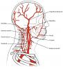
Sobotta 1909 fig.540 - Ramification of the external carotid artery in the head - English labels |
 |
Student128 |
Public Domain |
1.314 |
1 |
|
Data item |
MedicalGraphics - Drawing Bones of skull: anterior view - colour labels |
 |
rva |
Creative Commons Attribution-NoDerivatives |
1.313 |
4 |
|
Data item |
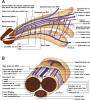
Gray - Drawing Fascial leg compartments in cross-section - English labels |
 |
rva |
Public Domain |
1.309 |
2 |
|
Data item |
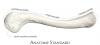
Anatomy Standard - Drawing Clavicula: superior view - Latin labels |
 |
rva |
Creative Commons Attribution-NonCommercial |
1.307 |
3 |
|
Anatomical Structure |
Incisura terminalis auricularis |
|
admin |
|
1.307 |
2 |
|
Learning Path |
Quiz Lever - Kenmerken en Onderdelen (gevorderd) |
|
tjscherphof |
Creative Commons Attribution-NonCommercial-ShareAlike |
1.307 |
0 |
|
Data item |
Slagter - Drawing Bones hand ventral side - no labels |
 |
lumcanatomy |
Creative Commons Attribution-NonCommercial-ShareAlike |
1.306 |
2 |
|
Data item |
Dundee - Drawing Superficial veins of the hand - English labels |
 |
rva |
Creative Commons Attribution-NonCommercial-NoDerivs |
1.305 |
3 |
|
Anatomical Structure |
Vertebrae cervicales (CI-CVII) |
|
admin |
|
1.303 |
2 |
|
Learning Path |
Quiz Thorax anatomy - clinical appliction |
|
EmmaL |
Creative Commons Attribution-NonCommercial-ShareAlike |
1.303 |
0 |
|
Data item |
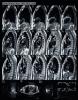
UBC - Website Horizontal Brain Slices - English labels |
|
M_Orsatti |
Creative Commons Attribution-NonCommercial-ShareAlike |
1.302 |
6 |
|
Data item |
Radiopaedia - Drawing Layers of the scalp and meninges - English labels |
 |
rva |
Creative Commons Attribution-NonCommercial-ShareAlike |
1.301 |
1 |
|
Data item |
BlueLink - 3D model Sixth Cervical Vertebra (C6) |
|
rva |
Creative Commons Attribution-NonCommercial-NoDerivs |
1.300 |
3 |
|
Data item |
Radiopaedia - Drawing Contents of superficial cubital fossa - English labels |
 |
rva |
Creative Commons Attribution-NonCommercial-ShareAlike |
1.299 |
3 |
|
Data item |
Anatomy Standard - Drawing Maxilla: posterior view - Latin labels |
 |
rva |
Creative Commons Attribution-NonCommercial |
1.298 |
2 |
|
Data item |
Leiden MOOC 5.6 - Video Anatomy on the table: demonstration of the superficial body wall |
|
opgobee |
Creative Commons Attribution-NonCommercial-ShareAlike |
1.298 |
4 |
|
Anatomical Structure |
Truncus coeliacus |
|
admin |
|
1.297 |
1 |
|
Data item |
U.Br.Columbia - Drawing Deep dissection of the sole of the foot - English labels |
 |
rva |
Creative Commons Attribution-NonCommercial-ShareAlike |
1.297 |
1 |
|
Data item |
RCSI - Drawing Superficial extensor muscles and tendons of forearm - English labels |
 |
rva |
Creative Commons Attribution-NonCommercial-ShareAlike |
1.296 |
3 |
|
Anatomical Structure |
Processus pterygoideus ossis sphenoidalis |
|
admin |
|
1.296 |
1 |
|
Learning Path |
Quiz Intestine - Vascularisation (basics) |
|
EmmaL |
Creative Commons Attribution-NonCommercial-ShareAlike |
1.296 |
1 |
|
Anatomical Structure |
Pars lateralis ossis sacri |
|
admin |
|
1.295 |
1 |
|
Data item |
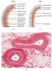
OpenStax AnatPhys fig.20.3 - Comparison of Artery and Vein - English labels |
 |
Jorn IJkhout |
Creative Commons Attribution |
1.295 |
3 |
|
Data item |
Sobotta 1909 fig.248 - cross section of the abdominal muscles, superior to linea arcuata - English labels |
 |
Student128 |
Public Domain |
1.294 |
1 |
|
Data item |
OpenStax AnatPhys fig.8.5 - Humerus and Elbow - English labels |
 |
Jorn IJkhout |
Creative Commons Attribution |
1.291 |
4 |
|
Data item |
Slagter - Drawing Upper abdominal organs with liver reflected superiorly - no labels |
 |
lumcanatomy |
Creative Commons Attribution-NonCommercial-ShareAlike |
1.290 |
3 |
|
Anatomical Structure |
Os temporale |
|
admin |
|
1.288 |
0 |
|
Learning Path |
Quiz Anatomie van Pijn C - Klinische toepassingen 1 |
|
opgobee |
Creative Commons Attribution-NonCommercial-ShareAlike |
1.288 |
2 |
|
Data item |
Radiopaedia - Drawing Anatomy of the rectum - English labels |
 |
rva |
Creative Commons Attribution-NonCommercial-ShareAlike |
1.287 |
3 |
|
Data item |
Anatomy Standard - Drawing Medial wall of the orbit - Latin labels |
 |
rva |
Creative Commons Attribution-NonCommercial |
1.286 |
3 |
|
Data item |
Anatomy Standard - Drawing Scapula: costal surface (anterior view) - Latin labels |
 |
rva |
Creative Commons Attribution-NonCommercial |
1.285 |
2 |
|
Anatomical Structure |
Lamina perpendicularis ossis ethmoidalis |
|
admin |
|
1.284 |
1 |
|
Data item |
Sobotta 1906 fig.508 - Uterus, tuba uterina, ovary and upper part of the vagina, frontal section - English labels |
 |
Student128 |
Public Domain |
1.282 |
3 |
|
Anatomical Structure |
Hepar |
|
admin |
|
1.281 |
3 |
|
Anatomical Structure |
Glandula suprarenalis |
|
admin |
|
1.280 |
2 |
|
Data item |
Sobotta 1909 fig.659 - Lamina quadrigemina and rhomboid fossa, posterior view - English labels |
 |
Student128 |
Public Domain |
1.280 |
3 |
|
Data item |
Dundee - Drawing Simplified Cross-Section of Heart - English labels |
 |
rva |
Creative Commons Attribution-NonCommercial-NoDerivs |
1.279 |
3 |
|
Data item |
Sobotta 1909 fig,100 - palate and maxilla, oral surface - English Labels |
 |
Student128 |
Public Domain |
1.278 |
1 |
|
Data item |
Radiopaedia - Drawing Bones of the knee joint: medial view - English labels |
 |
rva |
Creative Commons Attribution-NonCommercial-ShareAlike |
1.276 |
4 |
|
Data item |
OLI - Drawing Organs of the lympathic system - no labels |
 |
rva |
Creative Commons Attribution-NonCommercial-ShareAlike |
1.276 |
2 |
|
Data item |
3D Anatomy Lyon: The ligaments and vocal folds of the larynx - video of 3D model |
|
rva |
Creative Commons Attribution-NonCommercial-NoDerivs |
1.274 |
1 |
|
Data item |
Slagter - Drawing Abdominal organs in situ, anterior view - no labels |
") |
lumcanatomy |
Creative Commons Attribution-NonCommercial-ShareAlike |
1.273 |
2 |
|
Data item |
OpenStax AnatPhys fig.20.26 - Common Carotid Artery - English labels |
 |
Jorn IJkhout |
Creative Commons Attribution |
1.272 |
2 |
|
Anatomical Structure |
Os occipitale |
|
admin |
|
1.270 |
3 |
|
Data item |
Sobotta 1906 fig.459 - Transverse section of the thorax - English labels |
 |
Student128 |
Public Domain |
1.269 |
2 |
|
Anatomical Structure |
Facies articularis capitis costae |
|
admin |
|
1.268 |
3 |
|
Data item |
Rectocele - no labels |
 |
opgobee |
Creative Commons Attribution-NonCommercial-ShareAlike |
1.268 |
2 |
|
Data item |
Anatomy Standard - Drawing Maxilla: inferior view - Latin labels |
 |
rva |
Creative Commons Attribution-NonCommercial |
1.268 |
5 |
|
Anatomical Structure |
Sulci tendinum musculorum extensorum |
|
admin |
|
1.267 |
0 |
|
Learning Path |
Quiz Hart - Coronairvaten (gevorderd) |
|
opgobee |
Creative Commons Attribution-NonCommercial-ShareAlike |
1.267 |
0 |
|
Data item |
Radiopaedia - Drawing Right bronchial tree from posterior - English labels |
 |
rva |
Creative Commons Attribution-NonCommercial-ShareAlike |
1.266 |
3 |
|
Data item |
Lu - Drawing Vascularisation of penis: upper lateral view and cross-section - English labels |
 |
rva |
Creative Commons Attribution-NonCommercial-NoDerivs |
1.264 |
2 |
|
Data item |
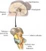
KnowledgeWorks - Drawing Pineal gland and thalamus - English labels |
 |
rva |
Creative Commons Attribution |
1.263 |
3 |
|
Data item |
Anatomy Standard - Drawing Apertura thoracis superior (thoracic inlet) - Latin labels |
 |
rva |
Creative Commons Attribution-NonCommercial |
1.262 |
6 |
|
Data item |
OpenStax AnatPhys fig.7.12 - Ethmoid Bone - English labels |
 |
Jorn IJkhout |
Creative Commons Attribution |
1.259 |
4 |
|
Data item |
Slagter - Drawing Glenohumeral (shoulder) joint: anterior view - no labels |
 |
rva |
Creative Commons Attribution-NonCommercial-ShareAlike |
1.258 |
3 |
|
Learning Path |
Demo quiz 2 - toont vraagmogelijkheden (NL) |
|
admin |
Creative Commons Attribution-NonCommercial-ShareAlike |
1.257 |
0 |
|
Data item |
OpenStax AnatPhys fig.22.12 -Lung Tissue - English labels |
 |
Jorn IJkhout |
Creative Commons Attribution |
1.257 |
2 |
|
Data item |
Radiopaedia - Drawing Aberrant right subclavian artery - English labels |
 |
rva |
Creative Commons Attribution-NonCommercial-NoDerivs |
1.257 |
3 |
|
Learning Path |
Quiz Hart - Ruimten (gevorderd) |
|
opgobee |
Creative Commons Attribution-NonCommercial-ShareAlike |
1.257 |
0 |
|
Data item |
Title convention AnatomyTOOL for Materials |
|
admin |
Creative Commons Attribution |
1.256 |
3 |
|
Data item |
Anatomy Standard - Drawing Ossa digitorum pedis: plantar view - no labels |
 |
rva |
Creative Commons Attribution-NonCommercial |
1.254 |
3 |
|
Anatomical Structure |
Os sphenoidale |
|
admin |
|
1.252 |
1 |
|
Anatomical Structure |
Intestinum tenue |
|
admin |
|
1.252 |
2 |
|
Data item |
Sobotta 1909 fig.632 - Fissures and convulsions of the cerebral cortex, medial surface - English labels |
 |
Student128 |
Public Domain |
1.252 |
4 |
|
Data item |
Universiteit Gent - Students - Drawing Hepar/Liver (2) - numbered Latin labels |
 |
sehellin |
Creative Commons Attribution-NonCommercial-ShareAlike |
1.251 |
4 |
|
Data item |
Anatomy Standard - Drawing Vertebral column: anterior, lateral and posterior view - no labels |
 |
rva |
Creative Commons Attribution-NonCommercial |
1.249 |
6 |
|
Data item |
Sobotta 1909 fig.237 - cross section of the back muscles - English labels |
 |
Student128 |
Public Domain |
1.248 |
5 |
|
Data item |
Leiden - Photo Opened duodenum with major duodenal papilla (dissection specimen) - no labels |
 |
opgobee |
Creative Commons Attribution-NonCommercial-ShareAlike |
1.248 |
2 |
|
Anatomical Structure |
Flexura coli dextra |
|
admin |
|
1.247 |
2 |
|
Anatomical Structure |
Circumferentia articularis capitis radii |
|
admin |
|
1.246 |
0 |
|
Data item |
RCSI - Drawing Arteries of kidney and adrenal gland - English labels |
 |
rva |
Creative Commons Attribution-NonCommercial-ShareAlike |
1.246 |
2 |
|
Data item |
Sobotta 1909 fig.251 - 12th rib and lumbal vertebra - English labels |
 |
Student128 |
Public Domain |
1.246 |
1 |
|
Data item |
Anatomy Standard - Drawing Third metacarpal bone - Latin labels |
 |
rva |
Creative Commons Attribution-NonCommercial |
1.244 |
2 |
|
Data item |
OpenStax AnatPhys fig.23.16 - Histology of Stomach - English labels |
 |
Jorn IJkhout |
Creative Commons Attribution |
1.244 |
1 |
|
Data item |
About Medicine: 3D model femoral triangle, femoral sheath and contained femoral vessels |
|
opgobee |
Creative Commons Attribution |
1.243 |
2 |
|
Data item |
Slagter - Different incisions for laparotomic approach pelvic surgery - no labels |
 |
Siem Zethof |
Creative Commons Attribution-NonCommercial-ShareAlike |
1.243 |
2 |
|
Data item |
Leiden - Drawing Difference in neurovascular connections superior and inferior of pectinate line - Latin and English labels |
 |
opgobee |
Creative Commons Attribution-ShareAlike |
1.242 |
3 |
|
Data item |
Slagter - Drawing Global anatomy of the brain: anterior view - Dutch labels |
 |
rva |
Creative Commons Attribution-NonCommercial-ShareAlike |
1.242 |
3 |
|
Data item |
CT thorax sagital |
 |
aherrler |
Creative Commons Attribution-NonCommercial-ShareAlike |
1.242 |
2 |
|
Data item |
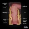
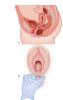
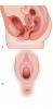
Cystocele - No labels |
 |
admin |
Creative Commons Attribution-NonCommercial-ShareAlike |
1.241 |
3 |
|
Data item |
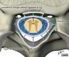
Radiopaedia - Drawing Ligamentum flavum, posterior longitudinal ligament and thecal sac - English labels |
 |
rva |
Creative Commons Attribution-NonCommercial-NoDerivs |
1.240 |
5 |
|
Data item |
OpenStax AnatPhys fig.15.4 - Connections of the Parasympathetic Nervous System - English labels |
 |
Jorn IJkhout |
Creative Commons Attribution |
1.240 |
2 |
|
Anatomical Structure |
Os ethmoidale |
|
admin |
|
1.239 |
3 |
|
Data item |
Radiopaedia - Drawing Contents and boundaries of the femoral triangle - English labels |
 |
rva |
Creative Commons Attribution-NonCommercial-ShareAlike |
1.239 |
1 |
|
Data item |
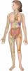
Drawing nervous system |
 |
lumcanatomy |
Creative Commons Attribution-NonCommercial-ShareAlike |
1.238 |
2 |
|
Data item |
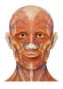
Lynch - Drawing Superficial anatomy of the head - no labels |
 |
rva |
Creative Commons Attribution |
1.238 |
8 |
|
Data item |
Anatomy Standard - Drawing Medial view of female pelvis with main pelvimetric conjugates - Latin labels |
 |
rva |
Creative Commons Attribution-NonCommercial |
1.236 |
2 |
|
Data item |
Anatomy Standard - Drawing Mandibula: superior view - Latin labels |
 |
rva |
Creative Commons Attribution-NonCommercial |
1.235 |
2 |
|
Anatomical Structure |
Bulbus oculi |
|
admin |
|
1.235 |
1 |
|
Data item |
Titel conventie AnatomyTOOL voor Materialen |
|
admin |
Creative Commons Attribution |
1.234 |
4 |
|
Data item |
3D Anatomy Lyon: The adductor muscles of the hip - video of 3D model |
|
rva |
Creative Commons Attribution-NonCommercial-NoDerivs |
1.233 |
2 |
|
Data item |
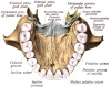
Anatomy Standard - Drawing Palatine bones: anterior view - Latin labels |
 |
rva |
Creative Commons Attribution-NonCommercial |
1.232 |
4 |
|
Learning Path |
Quiz Anatomie van Pijn B - Routes en Referred Pain 1 |
|
opgobee |
Creative Commons Attribution-NonCommercial-ShareAlike |
1.232 |
2 |
|
Data item |
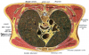
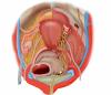
Superior view of the female pelvis with hypogastric plexus and internal iliac artery branches - no labels |
 |
Siem Zethof |
Creative Commons Attribution-NonCommercial-ShareAlike |
1.232 |
2 |
|
Anatomical Structure |
Facies orbitalis ossis frontalis |
|
admin |
|
1.228 |
2 |
|
Data item |
Sobotta 1909 fig.204 - articulations and ligaments of the hand, anterior view - English Labels |
 |
Student128 |
Public Domain |
1.227 |
1 |
|
Anatomical Structure |
Crista infratemporalis alaris majoris ossis sphenoidalis |
|
admin |
|
1.226 |
1 |
|
Anatomical Structure |
Collum glandis |
|
admin |
|
1.225 |
1 |