

nid: 58806
Additional formats:
None available
Description:
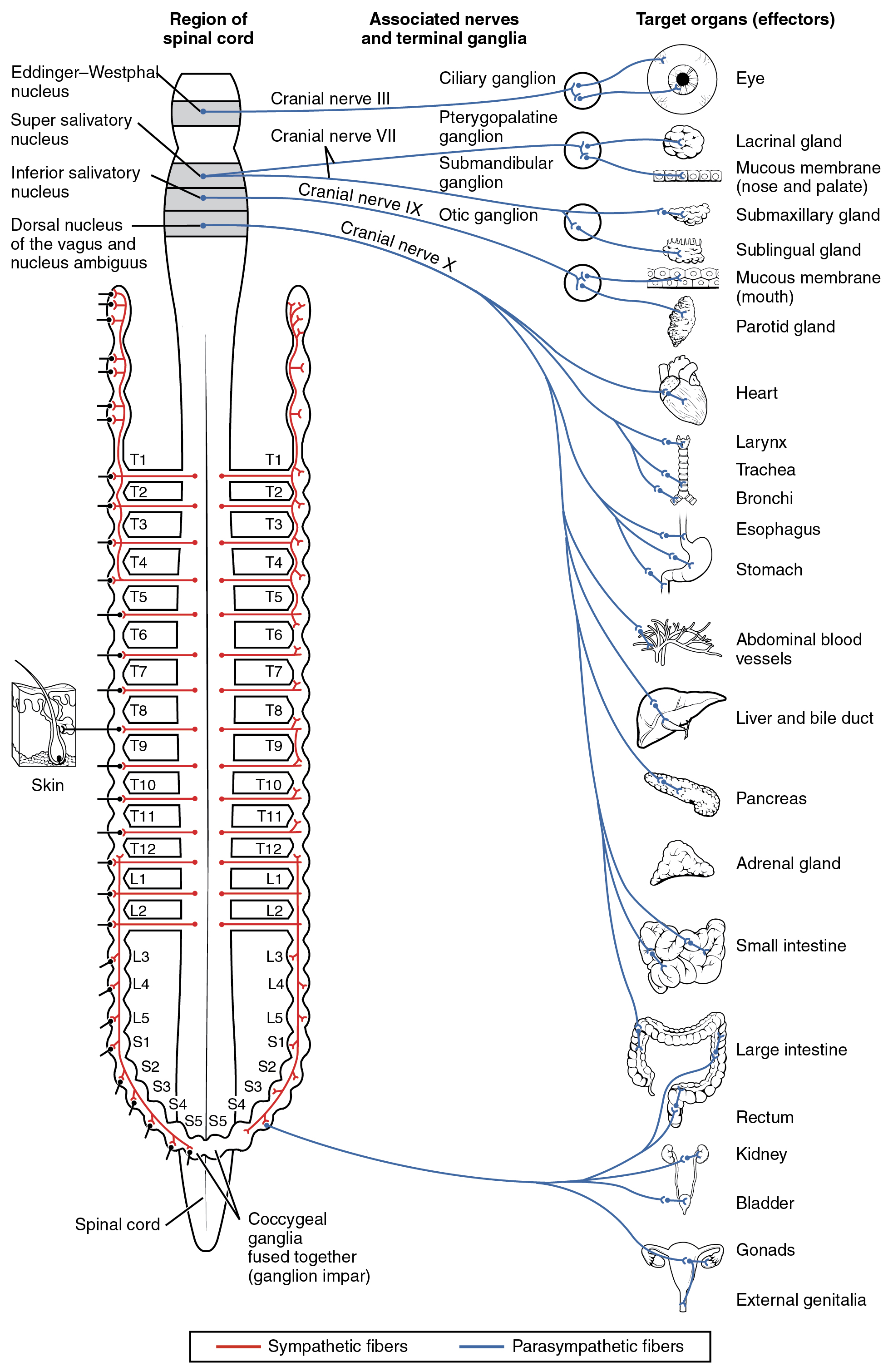
Connections of Parasympathetic Division of the Autonomic Nervous System Neurons from brainstem nuclei, or from the lateral horn of the sacral spinal cord, project to terminal ganglia near or within the various organs of the body. Axons from these ganglionic neurons then project the short distance to those target effectors. English labels. From OpenStax book 'Anatomy and Physiology', fig. 15.4.
Anatomical structures in item:
Uploaded by: Jorn IJkhout
Netherlands, Leiden – Leiden University Medical Center, Leiden University
Nervus
Pars parasympathica divisionis autonomici systematis nervosi
Ganglion parasympathicum
Medulla spinalis
Nucleus salivatorius superior
Nucleus salivatorius inferior
Nucleus dorsalis nervi vagi
Nucleus ambiguus
Nervus oculomotorius [III]
Nervus facialis [VII]
Nervus glossopharyngeus [IX]
Nervus vagus
Ganglion ciliare
Ganglion pterygopalatinum
Ganglion submandibulare
Ganglion oticum
Creator(s)/credit: OpenStax
Requirements for usage
You are free to use this item if you follow the requirements of the license:  View license
View license If you use this item you should credit it as follows:
- For usage in print - copy and paste the line below:
- For digital usage (e.g. in PowerPoint, Impress, Word, Writer) - copy and paste the line below (optionally add the license icon):
"OpenStax AnatPhys fig.15.4 - Connections of the Parasympathetic Nervous System - English labels" at AnatomyTOOL.org by OpenStax, license: Creative Commons Attribution. Source: book 'Anatomy and Physiology', https://openstax.org/details/books/anatomy-and-physiology.
"OpenStax AnatPhys fig.15.4 - Connections of the Parasympathetic Nervous System - English labels" by OpenStax, license: CC BY. Source: book 'Anatomy and Physiology', https://openstax.org/details/books/anatomy-and-physiology.


Comments