
nid: 60436
Additional formats:
None available
Description:
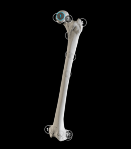
This 3D model shows the anatomy of the femur with annotations.
Anatomical structures in item:
Uploaded by: rva
Netherlands, Leiden – Leiden University Medical Center, Leiden University
Femur
Caput femoris
Collum femoris
Fovea capitis femoris
Trochanter major
Trochanter minor
Intertrochanteric crest of femur
Linea pectinea femoris
Tuberositas glutea
Linea aspera
Condylus medialis femoris
Epicondylus medialis femoris
Fossa intercondylaris
Condylus lateralis femoris
Epicondylus lateralis femoris
Creator(s)/credit: Dr Eric Bauer, Biology professor
Requirements for usage
You are free to use this item if you follow the requirements of the license:  View license
View license If you use this item you should credit it as follows:
- For usage in print - copy and paste the line below:
- For digital usage (e.g. in PowerPoint, Impress, Word, Writer) - copy and paste the line below (optionally add the license icon):
"Elon - 3D model Femur" at AnatomyTOOL.org by Eric Bauer, license: Creative Commons Attribution. Scanned and annotated by: students in Dr. Eric Bauer’s human anatomy lab at Elon University, North Carolina, USA.
"Elon - 3D model Femur" by Eric Bauer, license: CC BY. Scanned and annotated by: students in Dr. Eric Bauer’s human anatomy lab at Elon University, North Carolina, USA.


Comments