
nid: 60187
Additional formats:
None available
Description:
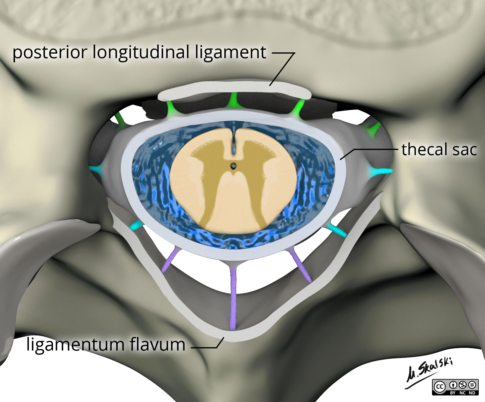
Ligamentum flavum, posterior longitudinal ligament and thecal sac. The anatomy of the ligamentum flavum, posterior longitudinal ligament and thecal sac (dural sac) can be seen in this image. English labels
Case courtesy of Dr Matt Skalski, Radiopaedia.org. From the case rID: 65838
Case courtesy of Dr Matt Skalski, Radiopaedia.org. From the case rID: 65838
Anatomical structures in item:
Uploaded by: rva
Netherlands, Leiden – Leiden University Medical Center, Leiden University
Spatium epidurale
Ligamentum longitudinale posterius
Ligamenta flava
Creator(s)/credit: Dr Matt Skalski MB.BS, MMed
Requirements for usage
You are free to use this item if you follow the requirements of the license:  View license
View license If you use this item you should credit it as follows:
- For usage in print - copy and paste the line below:
- For digital usage (e.g. in PowerPoint, Impress, Word, Writer) - copy and paste the line below (optionally add the license icon):
"Radiopaedia - Drawing Ligamentum flavum, posterior longitudinal ligament and thecal sac - English labels" at AnatomyTOOL.org by Matt Skalski, license: Creative Commons Attribution-NonCommercial-NoDerivs


Comments