|
Data item |
OpenStax AnatPhys fig.1.12 - Regions of the Human Body - English labels |
 |
opgobee |
Creative Commons Attribution |
1.969 |
1 |
|
Data item |
Cenveo - Drawing Structure and Function of the Semicircular Canals - English labels |
 |
rva |
Creative Commons Attribution |
1.966 |
3 |
|
Learning Path |
Quiz Thoracic wall (mixed subjects) 1 |
|
EmmaL |
Creative Commons Attribution-NonCommercial-ShareAlike |
1.966 |
4 |
|
Data item |
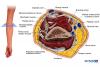
OpenStax AnatPhys fig.20.33 - Lower Limb Arteries Anterior Posterior - English labels |
 |
Jorn IJkhout |
Creative Commons Attribution |
1.964 |
3 |
|
Anatomical Structure |
Angulus costae |
|
admin |
|
1.964 |
0 |
|
Data item |
Evaluatierapporten project AnatomyTOOL |
|
lumcanatomy |
Creative Commons Attribution |
1.960 |
1 |
|
Anatomical Structure |
Dorsal tubercle of radius |
|
admin |
|
1.959 |
2 |
|
Data item |
Lynch - Drawing Diagram of heart in lateral view - English labels |
 |
rva |
Creative Commons Attribution |
1.959 |
7 |
|
Data item |
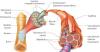
Vascularisation gut (exploded view) |
") |
lumcanatomy |
Creative Commons Attribution-NonCommercial-ShareAlike |
1.957 |
2 |
|
Data item |
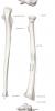
Anatomy Standard - Drawing Proximal ulna and radius - Latin labels |
 |
rva |
Creative Commons Attribution-NonCommercial |
1.956 |
4 |
|
Data item |
3D Anatomy Lyon: Anatomy of the shoulder - video of 3D model |
|
rva |
Creative Commons Attribution-NonCommercial-NoDerivs |
1.956 |
3 |
|
Data item |
Rotation video of 3D reconstruction female pelvis, pelvic diaphragm and organs - build, no labels |
|
opgobee |
Creative Commons Attribution-NonCommercial-ShareAlike |
1.955 |
1 |
|
Data item |
NYSORA - Drawing Cross-section of elbow - English labels |
 |
rva |
Creative Commons Attribution-NonCommercial-NoDerivs |
1.953 |
2 |
|
Anatomical Structure |
Margo lateralis scapulae |
|
admin |
|
1.950 |
1 |
|
Data item |
Leiden - Drawing Taenia libera and taenia omentalis - Latin labels |
 |
opgobee |
Creative Commons Attribution-NonCommercial-ShareAlike |
1.950 |
4 |
|
Data item |
Lateral view of opened thorax pericardium opened |
 |
Nadja Baltensweiler |
Creative Commons Attribution-NonCommercial-ShareAlike |
1.948 |
3 |
|
Data item |
RCSI - Drawing Circle of Willis - English labels |
 |
rva |
Creative Commons Attribution-NonCommercial-ShareAlike |
1.947 |
1 |
|
Data item |
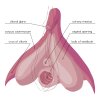
Amphis - Drawing Internal anatomy of the clitoris - English labels |
 |
rva |
Public Domain |
1.946 |
4 |
|
Anatomical Structure |
Ostium ureteris |
|
admin |
|
1.937 |
1 |
|
Data item |
Leiden - E-learning CASK Inguinal Area |
|
opgobee |
This item is (on) an external site. The license as stated on that site holds. |
1.930 |
2 |
|
Anatomical Structure |
Tuberositas masseterica |
|
admin |
|
1.927 |
1 |
|
Data item |
OpenStax AnatPhys fig.9.12 - Body Movements(Page 1) - English labels |
 |
Jorn IJkhout |
Creative Commons Attribution |
1.926 |
3 |
|
Data item |
3D Anatomy Lyon: Anatomy of the teres major - video of 3D model |
|
rva |
Creative Commons Attribution-NonCommercial-NoDerivs |
1.924 |
1 |
|
Data item |
Sobotta 1909 fig.628 - Fissures and convulsions of the cerebral cortex, superior view - English labels |
 |
Student128 |
Public Domain |
1.923 |
1 |
|
Data item |
Radiopaedia - Drawing Left bronchial tree from anterior - English labels |
 |
rva |
Creative Commons Attribution-NonCommercial-NoDerivs |
1.920 |
9 |
|
Data item |
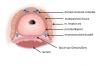
Dundee - Drawing Anatomy of the ear - English labels |
 |
rva |
Creative Commons Attribution-NonCommercial-NoDerivs |
1.920 |
3 |
|
Data item |
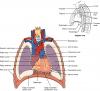
OpenStax AnatPhys fig.19.2 - Heart Position in Thorax - English labels |
 |
Jorn IJkhout |
Creative Commons Attribution |
1.917 |
5 |
|
Learning Path |
Quiz Buikorganen - Vascularisatie (basis) |
|
opgobee |
Creative Commons Attribution-NonCommercial-ShareAlike |
1.917 |
0 |
|
Learning Path |
Quiz Liver - Vascularisation (advanced) |
|
EmmaL |
Creative Commons Attribution-NonCommercial-ShareAlike |
1.916 |
1 |
|
Anatomical Structure |
Promontorium ossis sacri |
|
admin |
|
1.916 |
1 |
|
Data item |
Anatomy Standard - Drawing Basis cranii externa: detail of openings - Latin labels |
 |
rva |
Creative Commons Attribution-NonCommercial |
1.914 |
1 |
|
Data item |
Anatomy Standard - Drawing Scapula: lateral view - Latin labels |
 |
rva |
Creative Commons Attribution-NonCommercial |
1.914 |
6 |
|
Data item |
Elon - 3D model Radius |
|
rva |
Creative Commons Attribution |
1.912 |
2 |
|
Learning Path |
Quiz Lever - Vascularisatie (gevorderd) |
|
tjscherphof |
Creative Commons Attribution-NonCommercial-ShareAlike |
1.911 |
0 |
|
Data item |
Anatomy Standard - Drawing Maxilla: lateral view - Latin labels |
 |
rva |
Creative Commons Attribution-NonCommercial |
1.906 |
4 |
|
Data item |
Sobotta 1909 fig.39 - temporal lines of the skull - English Labels |
 |
Student128 |
Public Domain |
1.900 |
4 |
|
Data item |
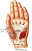
U.Br.Columbia - Drawing Superficial dissection of the palm - English labels |
 |
rva |
Creative Commons Attribution-NonCommercial-ShareAlike |
1.896 |
1 |
|
Data item |
U.Br.Columbia - Drawing Innervation of the parotid gland - English labels |
 |
rva |
Creative Commons Attribution-NonCommercial-ShareAlike |
1.894 |
2 |
|
Data item |
OpenStax AnatPhys fig.8.14 - Ligaments of Pelvis - no labels |
 |
rva |
Creative Commons Attribution |
1.888 |
2 |
|
Data item |
Bladder control and micturition |
|
opgobee |
Creative Commons Attribution-NonCommercial-ShareAlike |
1.877 |
4 |
|
Data item |
Leiden - Drawing Muscles And Erectile Bodies In Female Perineum - English labels |
 |
opgobee |
Creative Commons Attribution-NonCommercial-ShareAlike |
1.875 |
2 |
|
Learning Path |
BBS1002 Human anatomy of the cardiovascular system |
|
leo.koehler |
Creative Commons Attribution-NonCommercial-ShareAlike |
1.873 |
0 |
|
Data item |
Anatomy Standard - Drawing Right radius: posterior view - Latin labels |
 |
rva |
Creative Commons Attribution-NonCommercial |
1.872 |
2 |
|
Data item |
Sobotta 1909 fig.660 - Lamina quadrigemina and rhomboid fossa, posterior view - English labels |
 |
Student128 |
Public Domain |
1.872 |
4 |
|
Data item |
Anatomy Standard - Drawing Basis cranii interna - Latin labels |
 |
rva |
Creative Commons Attribution-NonCommercial |
1.871 |
2 |
|
Data item |
U.Br.Columbia - Drawing Overview of the brachial plexus - no labels |
 |
rva |
Creative Commons Attribution-NonCommercial-ShareAlike |
1.869 |
3 |
|
Anatomical Structure |
Fovea dentis |
|
admin |
|
1.863 |
1 |
|
Data item |
Anatomy Standard - Drawing Lacrimal bone: lateral view - Latin labels |
 |
rva |
Creative Commons Attribution-NonCommercial |
1.860 |
8 |
|
Data item |
Anatomy Standard - Drawing Maxilla: medial view - Latin labels |
 |
rva |
Creative Commons Attribution-NonCommercial |
1.859 |
4 |
|
Learning Path |
Quiz Inwendige genitaliën Vrouw - Vascularisatie (basis) |
|
tjscherphof |
Creative Commons Attribution-NonCommercial-ShareAlike |
1.859 |
0 |
|
Data item |
Blausen 0451 - Anterior view of the heart - English labels |
 |
Student10 |
Creative Commons Attribution |
1.858 |
3 |
|
Data item |
Vascularisation gut |
 |
lumcanatomy |
Creative Commons Attribution-NonCommercial-ShareAlike |
1.858 |
1 |
|
Data item |
Anatomy Standard - Drawing Ligamentum cruciforme atlantis: dorsal view - Latin labels |
 |
rva |
Creative Commons Attribution-NonCommercial |
1.856 |
2 |
|
Data item |
U.Br.Columbia - Drawing Deep anatomy of the popliteal fossa - English labels |
 |
rva |
Creative Commons Attribution-NonCommercial-ShareAlike |
1.851 |
1 |
|
Data item |
OpenStax AnatPhys fig.22.7 - The Larynx - English labels |
 |
Jorn IJkhout |
Creative Commons Attribution |
1.849 |
2 |
|
Data item |
U.Br.Columbia - Drawing Origins and insertions of the pharyngeal muscles - English labels |
 |
rva |
Creative Commons Attribution-NonCommercial-ShareAlike |
1.845 |
2 |
|
Data item |
Tempel - Drawing Lateral view of larynx: superior laryngeal nerve - English labels |
 |
rva |
Creative Commons Attribution-NonCommercial-NoDerivs |
1.840 |
5 |
|
Data item |
Anatomy Standard - Drawing Basis cranii interna: dorsolateral view - Latin labels |
 |
rva |
Creative Commons Attribution-NonCommercial |
1.837 |
2 |
|
Data item |
BlueLink - 3D model Second right rib |
|
rva |
Creative Commons Attribution-NonCommercial-NoDerivs |
1.837 |
12 |
|
Data item |
Sobotta 1909 fig.249 - cross section of the abdominal muscles, inferior to linea arcuata - English labels |
 |
Student128 |
Public Domain |
1.836 |
2 |
|
Learning Path |
SIP: BBS1005: Case 1- Should we worry? + DNA practical |
|
Tatiana Sviriniuc |
Creative Commons Attribution-NonCommercial-ShareAlike |
1.834 |
0 |
|
Data item |
Leiden - Drawing Cross-section upper abdomen with peritoneal lining - no labels |
") |
opgobee |
Creative Commons Attribution-NonCommercial-ShareAlike |
1.834 |
2 |
|
Data item |
Thunthu - 3D model Pelvic bone |
|
rva |
Creative Commons Attribution |
1.830 |
1 |
|
Data item |
Sobotta 1909 fig.37 - The skull, anterior view - No labels |
 |
Student128 |
Public Domain |
1.829 |
3 |
|
Data item |
Slagter - Drawing Male inguinal area internal view - no labels |
 |
opgobee |
Creative Commons Attribution-NonCommercial-ShareAlike |
1.829 |
1 |
|
Data item |
OLI - Drawing Structure of skeletal muscle and sheats - English labels |
 |
rva |
Creative Commons Attribution-NonCommercial-ShareAlike |
1.820 |
3 |
|
Data item |
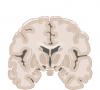
Slagter - Drawing Coronal section of the brain - no labels |
 |
rva |
Creative Commons Attribution-NonCommercial-ShareAlike |
1.814 |
3 |
|
Data item |
Groningen - 3D model Heart with Left Dominance - numbered English and Latin labels |
|
rva |
Creative Commons Attribution-NonCommercial-ShareAlike |
1.804 |
3 |
|
Data item |
Anatomy Standard - Drawing Temporal fossa - Latin labels |
 |
rva |
Creative Commons Attribution-NonCommercial |
1.802 |
4 |
|
Data item |
Radiopaedia - Drawing Midbrain at level of superior colliculus and oculomotor nerve - English labels |
 |
rva |
Creative Commons Attribution-NonCommercial-ShareAlike |
1.801 |
3 |
|
Data item |
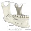
Anatomy Standard - Drawing Mandibula: anterolateral view - Latin labels |
 |
rva |
Creative Commons Attribution-NonCommercial |
1.799 |
5 |
|
Data item |
Sobotta 1909 fig.282 - aponeurosis palmaris - English Labels |
 |
Student128 |
Public Domain |
1.795 |
1 |
|
Data item |
U.Br.Columbia - Drawing The middle mediastinum - English labels |
 |
rva |
Creative Commons Attribution-NonCommercial-ShareAlike |
1.787 |
8 |
|
Data item |
Leiden - Video Colonoscopy: transition from ileum to caecum - English labels |
|
opgobee |
Creative Commons Attribution-NonCommercial-ShareAlike |
1.783 |
2 |
|
Data item |
RCSI - Drawing Cross-section of the knee joint - English labels |
 |
rva |
Creative Commons Attribution-NonCommercial-ShareAlike |
1.779 |
2 |
|
Data item |
Slagter - Drawing Transverse section of prostate, fascias and levator ani - Dutch labels |
 |
Siem Zethof |
Creative Commons Attribution-NonCommercial-ShareAlike |
1.778 |
1 |
|
Data item |
Leiden - Drawing Surface projection of digestive tract on thoracic cage (transparent organs) - no labels |
") |
lumcanatomy |
Creative Commons Attribution-NonCommercial-ShareAlike |
1.773 |
3 |
|
Data item |
U.Br.Columbia - Drawing Origins and insertions of the laryngeal muscles - English labels |
 |
rva |
Creative Commons Attribution-NonCommercial-ShareAlike |
1.772 |
1 |
|
Learning Path |
Quiz Inwendige Genitaliën Vrouw (gevorderd) |
|
tjscherphof |
Creative Commons Attribution-NonCommercial-ShareAlike |
1.768 |
0 |
|
Data item |
Sobotta 1909 fig.137 - femur, posterior view - colour, no labels |
 |
opgobee |
Creative Commons Attribution-ShareAlike |
1.767 |
1 |
|
Data item |
Lynch - Drawing Apical four-chamber diagram of heart - English labels |
 |
rva |
Creative Commons Attribution |
1.766 |
3 |
|
Data item |
OpenStax AnatPhys fig.7.13 - Nasal Cavity - English labels |
 |
Jorn IJkhout |
Creative Commons Attribution |
1.765 |
9 |
|
Anatomical Structure |
Apertura pelvis superior |
|
admin |
|
1.764 |
2 |
|
Data item |
Blausen 0896 - Ventricular system of the brain - English labels |
 |
Student10 |
Creative Commons Attribution |
1.763 |
1 |
|
Data item |
TEMPLATE Anatomical Topics TOC |
|
lumctest |
Creative Commons Attribution-NonCommercial-ShareAlike |
1.761 |
1 |
|
Data item |
Abdominal wall and pelvic floor in breathing and increased abdominal pressure |
|
opgobee |
Creative Commons Attribution-NonCommercial-ShareAlike |
1.761 |
2 |
|
Anatomical Structure |
Plica glossoepiglottica mediana |
|
admin |
|
1.759 |
0 |
|
Data item |
OpenStax AnatPhys fig.13.23 - The Cranial Nerves - English labels |
 |
Jorn IJkhout |
Creative Commons Attribution |
1.759 |
1 |
|
Data item |
Anatomy Standard - Drawing Sphenoid bone: inferior view - Latin labels |
 |
rva |
Creative Commons Attribution-NonCommercial |
1.759 |
4 |
|
Data item |
Leiden - Presentation slides Inguinal area, internal view, and entry points of inguinal and femoral hernia’s |
|
opgobee |
Creative Commons Attribution-NonCommercial-ShareAlike |
1.759 |
3 |
|
Anatomical Structure |
Arcus zygomaticus |
|
admin |
|
1.758 |
4 |
|
Data item |
Anatomy Standard - Drawing Sinus tarsi, sulcus tendinis, and trochlea peronealis - Latin labels |
 |
rva |
Creative Commons Attribution-NonCommercial |
1.758 |
2 |
|
Data item |
Servier - Drawing Brain sagittal section - no labels |
 |
rva |
Creative Commons Attribution |
1.758 |
8 |
|
Data item |
Anatomy Standard - Drawing Ligamentum capitis costae intra-articulare - Latin labels |
 |
rva |
Creative Commons Attribution-NonCommercial |
1.756 |
5 |
|
Data item |
Slagter - Drawing Human spermatogenesis diagram - English labels |
 |
rva |
Creative Commons Attribution-NonCommercial-ShareAlike |
1.756 |
5 |
|
Data item |
OpenStax AnatPhys fig.20.32 - Thoracic Upper Limb Arteries Chart - English labels |
 |
Jorn IJkhout |
Creative Commons Attribution |
1.746 |
3 |
|
Data item |
Anatomy Standard - Drawing Phalanx proximalis, media and distalis pedis: dorsal, plantar, and medial view - Latin labels |
 |
rva |
Creative Commons Attribution-NonCommercial |
1.746 |
5 |
|
Data item |
Leiden - Drawing Calot's triangle - Latin labels |
 |
opgobee |
Creative Commons Attribution-NonCommercial-ShareAlike |
1.740 |
2 |
|
Anatomical Structure |
Trigonum vesicae |
|
admin |
|
1.738 |
2 |
|
Anatomical Structure |
Canalis analis |
|
admin |
|
1.736 |
1 |