
nid: 63114
Additional formats:
None available
Description:
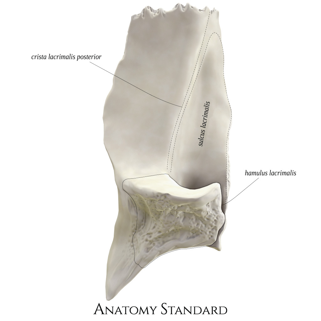
Lacrimal bone: lateral view. The lacrimal bones (os lacrimale) border the nasolacrimal canal. This image shows the right lacrimal bone from lateral. The bone is located between the frontal process of maxilla and the orbital plate of ethmoid. Latin labels.
Image retrieved from Anatomy Standard.
Image retrieved from Anatomy Standard.
Anatomical structures in item:
Uploaded by: rva
Netherlands, Leiden – Leiden University Medical Center, Leiden University
Os lacrimale
Crista lacrimalis posterior
Sulcus lacrimalis
Hamulus lacrimalis
Creator(s)/credit: Jānis Šavlovskis MD, PhD, Assistant Professor; Kristaps Raits, 3D generalist
Requirements for usage
You are free to use this item if you follow the requirements of the license:  View license
View license If you use this item you should credit it as follows:
- For usage in print - copy and paste the line below:
- For digital usage (e.g. in PowerPoint, Impress, Word, Writer) - copy and paste the line below (optionally add the license icon):
"Anatomy Standard - Drawing Lacrimal bone: lateral view - Latin labels" at AnatomyTOOL.org by Jānis Šavlovskis and Kristaps Raits, license: Creative Commons Attribution-NonCommercial


Comments