|
Data item |
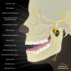
Radiopaedia - Drawing Main branches of the mandibular nerve - English labels |
 |
rva |
Creative Commons Attribution-NonCommercial-ShareAlike |
2.287 |
2 |
|
Data item |
Rotation video of 3D reconstruction female pelvis, pelvic diaphragm and organs - complete, no labels |
|
opgobee |
Creative Commons Attribution-NonCommercial-ShareAlike |
2.283 |
2 |
|
Data item |
Leiden-Delft - 3D model UAH viewer pelvis |
|
opgobee |
Creative Commons Attribution-NonCommercial-ShareAlike |
2.283 |
2 |
|
Data item |
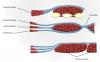
Slagter - Drawing Spermatogenesis in seminiferous tubules of testis - English labels |
 |
rva |
Creative Commons Attribution-NonCommercial-ShareAlike |
2.279 |
4 |
|
Learning Path |
Quiz Lungs - Pleurae and Recesses |
|
EmmaL |
Creative Commons Attribution-NonCommercial-ShareAlike |
2.278 |
2 |
|
Data item |
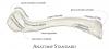
Anatomy Standard - Drawing Right radius: anterior view - Latin labels |
 |
rva |
Creative Commons Attribution-NonCommercial |
2.277 |
7 |
|
Data item |
MedicalGraphics - Drawing Larynx: lateral view and sagittal section - English labels |
 |
rva |
Creative Commons Attribution-NoDerivatives |
2.275 |
4 |
|
Learning Path |
Quiz hart (gemengde onderwerpen) 1 |
|
opgobee |
Creative Commons Attribution-NonCommercial-ShareAlike |
2.262 |
3 |
|
Data item |
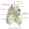
Anatomy Standard - Drawing Ethmoid bone: superior view - Latin labels |
 |
rva |
Creative Commons Attribution-NonCommercial |
2.259 |
7 |
|
Learning Path |
Quiz Dikke Darm - Kenmerken en Onderdelen (gevorderd) |
|
tjscherphof |
Creative Commons Attribution-NonCommercial-ShareAlike |
2.255 |
0 |
|
Data item |
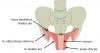
Schematic view of autonomic nerves, and visceral and somatic afferent nerves of the male pelvic organs – Dutch labels |
 |
Siem Zethof |
Creative Commons Attribution-NonCommercial-ShareAlike |
2.254 |
1 |
|
Data item |
Utah - Website NeuroLogic Exam |
|
M_Orsatti |
Creative Commons Attribution-NonCommercial-ShareAlike |
2.253 |
4 |
|
Data item |
U.Br.Columbia - Drawing The brachial plexus - English labels |
 |
rva |
Creative Commons Attribution-ShareAlike |
2.251 |
1 |
|
Anatomical Structure |
Facies articularis superior vertebrae |
|
admin |
|
2.247 |
0 |
|
Data item |
NYSORA - Drawing Structures of upper thigh and pelvis - English labels |
 |
rva |
Creative Commons Attribution-NonCommercial-NoDerivs |
2.240 |
3 |
|
Data item |
Anatomy Standard - Drawing Clavicula: inferior view - Latin labels |
 |
rva |
Creative Commons Attribution-NonCommercial |
2.239 |
5 |
|
Data item |
Cenveo - Drawing Anatomy of long bones - no labels |
 |
rva |
Creative Commons Attribution |
2.230 |
9 |
|
Data item |
Sobotta 1906 fig.511 - Female erectile structures and greater vestibular glands - English labels |
 |
Student128 |
Public Domain |
2.229 |
6 |
|
Data item |
Anatomy Standard - Drawing Infratemporal fossa: lateroinferior view - Latin labels |
 |
rva |
Creative Commons Attribution-NonCommercial |
2.223 |
3 |
|
Data item |
Anatomy Standard - Drawing Occipital bone: exterior (posterior) view - Latin labels |
 |
rva |
Creative Commons Attribution-NonCommercial |
2.221 |
2 |
|
Data item |
Anterior view of funnel-shaped attachement of levator ani to the obturator internus muscle – Dutch labels |
 |
Siem Zethof |
Creative Commons Attribution-NonCommercial-ShareAlike |
2.218 |
5 |
|
Data item |
Sobotta 1906 fig.423 - Lateral wall of the nasal cavity - English labels |
 |
Student128 |
Public Domain |
2.215 |
1 |
|
Data item |
Slagter - Drawing Bones and joints of foot: anterior and lateral view - Latin labels |
 |
rva |
Creative Commons Attribution-NonCommercial-ShareAlike |
2.215 |
1 |
|
Anatomical Structure |
Crista sacralis mediana |
|
admin |
|
2.214 |
0 |
|
Learning Path |
Quiz Peritoneum - Peritoneale relaties buikorganen (basis) |
|
opgobee |
Creative Commons Attribution-NonCommercial-ShareAlike |
2.213 |
0 |
|
Data item |
Hepatic segmentation |
 |
lumcanatomy |
Creative Commons Attribution-ShareAlike |
2.210 |
4 |
|
Data item |
Dundee - Drawing Anatomy of the Inner Ear - English labels |
 |
rva |
Creative Commons Attribution-NonCommercial-NoDerivs |
2.203 |
2 |
|
Data item |
Thunthu - 3D model Bones of the foot |
|
rva |
Creative Commons Attribution |
2.198 |
1 |
|
Data item |
Anatomy Standard - Drawing Humerus: posterior view - no labels |
 |
rva |
Creative Commons Attribution-NonCommercial |
2.191 |
18 |
|
Data item |
RCSI - Drawing Rectus sheath above and below arcuate line - English labels |
 |
rva |
Creative Commons Attribution-NonCommercial-ShareAlike |
2.191 |
2 |
|
Data item |
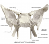
Anatomy Standard - Drawing Sphenoid bone: posterior view - Latin labels |
 |
rva |
Creative Commons Attribution-NonCommercial |
2.178 |
8 |
|
Data item |
Jmarchn - Drawing Human Anus - no labels |
 |
opgobee |
Creative Commons Attribution-ShareAlike |
2.175 |
7 |
|
Data item |
3D Anatomy Lyon: Muscles for abduction of the arm - video of 3D model |
|
rva |
Creative Commons Attribution-NonCommercial-NoDerivs |
2.166 |
2 |
|
Data item |
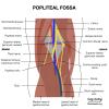
Radiopaedia - Drawing Contents of the popliteal fossa - English labels |
 |
rva |
Creative Commons Attribution-NonCommercial-ShareAlike |
2.165 |
1 |
|
Data item |
Anatomy Standard - Drawing Calcaneus bone in situ & ex situ: medial view - Latin labels |
 |
rva |
Creative Commons Attribution-NonCommercial |
2.158 |
5 |
|
Data item |
Human Biology fig. 1.66 - Overview of the organs of the digestive system - English labels |
 |
rva |
Creative Commons Attribution-NonCommercial |
2.157 |
3 |
|
Learning Path |
Quiz Dunne Darm - Kenmerken en Onderdelen (gevorderd) |
|
tjscherphof |
Creative Commons Attribution-NonCommercial-ShareAlike |
2.154 |
0 |
|
Data item |
Slagter - Drawing Anatomy of the penis and corpus cavernosum - no labels |
 |
rva |
Creative Commons Attribution-NonCommercial-ShareAlike |
2.147 |
5 |
|
Anatomical Structure |
Os costale (Costa) |
|
admin |
|
2.146 |
1 |
|
Data item |
OpenStax AnatPhys fig.1.15 - Dorsal and Ventral Body Cavities - English labels |
 |
Jorn IJkhout |
Creative Commons Attribution |
2.139 |
4 |
|
Data item |
Urine and bowel continence |
|
opgobee |
Creative Commons Attribution-NonCommercial-ShareAlike |
2.138 |
2 |
|
Data item |
Anatomy Standard - Drawing Anatomy of the acetabulum - Latin labels |
 |
rva |
Creative Commons Attribution-NonCommercial |
2.136 |
2 |
|
Data item |
Anatomy Standard - Drawing Scapula: posterior surface - Latin labels |
 |
rva |
Creative Commons Attribution-NonCommercial |
2.135 |
3 |
|
Data item |
Leiden - Video Drie lokaties organen ten opzichte van het peritoneum |
|
opgobee |
Creative Commons Attribution-NonCommercial-ShareAlike |
2.134 |
4 |
|
Data item |
3D Anatomy Lyon: The sternoclavicular joint - video of 3D model |
|
rva |
Creative Commons Attribution-NonCommercial-NoDerivs |
2.131 |
3 |
|
Anatomical Structure |
Sulcus nervi spinalis |
|
admin |
|
2.121 |
2 |
|
Data item |
OpenStax AnatPhys fig.11.5 - Anterior and Posterior Views of Muscles - English labels |
 |
Jorn IJkhout |
Creative Commons Attribution |
2.116 |
3 |
|
Data item |
Cenveo - Drawing Larynx and vocal cords - English labels |
 |
rva |
Creative Commons Attribution |
2.112 |
1 |
|
Data item |
Elon - 3D model Sacrum and Coccyx |
|
rva |
Creative Commons Attribution |
2.110 |
7 |
|
Learning Path |
Quiz Maag - Kenmerken en Onderdelen (gevorderd) |
|
tjscherphof |
Creative Commons Attribution-NonCommercial-ShareAlike |
2.106 |
0 |
|
Data item |
OpenStax AnatPhys fig.6.12a - Compact Bone - English labels |
 |
Jorn IJkhout |
Creative Commons Attribution |
2.105 |
1 |
|
Data item |
Fribourg, Lausanne, Bern - Website Embryology.ch |
|
opgobee |
This item is (on) an external site. The license as stated on that site holds. |
2.097 |
4 |
|
Data item |
Slagter - Drawing View into omental bursa - no labels |
 |
lumcanatomy |
Creative Commons Attribution-NonCommercial-ShareAlike |
2.096 |
1 |
|
Data item |
OpenStax AnatPhys fig.20.40 - FlowChart Veins into VenaCava - English labels |
 |
Jorn IJkhout |
Creative Commons Attribution |
2.093 |
4 |
|
Learning Path |
Quiz Lungs - Characteristics and Parts (advanced) |
|
EmmaL |
Creative Commons Attribution-NonCommercial-ShareAlike |
2.093 |
0 |
|
Data item |
Radiopaedia - Drawing Scapula lateral view - English labels |
 |
rva |
Creative Commons Attribution-NonCommercial-NoDerivs |
2.092 |
12 |
|
Data item |
Servier - Drawing Vertebral column posterior view - no labels |
 |
rva |
Creative Commons Attribution |
2.091 |
2 |
|
Data item |
Blausen 0400 - Female reproductive system (Lateral view) - English labels |
 |
Student10 |
Creative Commons Attribution |
2.089 |
27 |
|
Data item |
Leiden - Drawing Cross-section upper abdomen with peritoneal lining - English labels |
 |
admin |
Creative Commons Attribution-NonCommercial-ShareAlike |
2.088 |
5 |
|
Data item |
Tendinous Arch of Levator Ani, Tendinous Arch Of Pelvic Fascia And Ligaments Of Female Pelvis - no labels |
 |
admin |
Creative Commons Attribution-NonCommercial-ShareAlike |
2.085 |
2 |
|
Data item |
Slagter -Drawing Peritoneal reflections on the female internal pelvic organs - English labels |
 |
opgobee |
Creative Commons Attribution-NonCommercial-ShareAlike |
2.080 |
8 |
|
Data item |
Slagter - Drawing Fetal circulation - no labels |
 |
opgobee |
Creative Commons Attribution-NonCommercial-ShareAlike |
2.079 |
3 |
|
Data item |
Superior view of female pelvis viscera, peritoneum, arterial supply and pelvic plexus – Dutch labels |
 |
Siem Zethof |
Creative Commons Attribution-NonCommercial-ShareAlike |
2.079 |
2 |
|
Data item |
OpenStax AnatPhys fig.20.24 - Major Systemic Artery - English labels |
 |
Jorn IJkhout |
Creative Commons Attribution |
2.076 |
11 |
|
Anatomical Structure |
Tuberositas pronatoria |
|
admin |
|
2.067 |
2 |
|
Learning Path |
Quiz Thoracic Wall - Skeleton Characteristics and parts |
|
EmmaL |
Creative Commons Attribution-NonCommercial-ShareAlike |
2.065 |
0 |
|
Data item |
U.Br.Columbia - Drawing Origins and insertions of the intrinsic tongue muscles - English labels |
 |
rva |
Creative Commons Attribution-NonCommercial-ShareAlike |
2.064 |
1 |
|
Data item |
Anatomy Standard - Drawing Sacrum: anterior aspect - Latin labels |
 |
rva |
Creative Commons Attribution-NonCommercial |
2.062 |
4 |
|
Data item |
Anatomy Standard - Drawing Knee joint and patella: anterior, medial and posterior view - no labels |
 |
rva |
Creative Commons Attribution-NonCommercial |
2.060 |
8 |
|
Anatomical Structure |
Costa prima [I] |
|
admin |
|
2.053 |
1 |
|
Data item |
Slagter - Drawing Radius and ulna separate and with interosseous membrane - no labels |
 |
lumcanatomy |
Creative Commons Attribution-NonCommercial-ShareAlike |
2.051 |
4 |
|
Anatomical Structure |
Lamina horizontalis ossis palatini |
|
admin |
|
2.049 |
1 |
|
Data item |
Dundee - Drawing Glossopharyngeal nerve and referred otalgia - English labels |
 |
rva |
Creative Commons Attribution-NonCommercial-NoDerivs |
2.049 |
1 |
|
Data item |
Vallance - 3D model Female reproductive organs |
|
rva |
Creative Commons Attribution |
2.048 |
1 |
|
Data item |
Sobotta 1909 fig.255 - neck muscles - no labels |
 |
rva |
Creative Commons Attribution-ShareAlike |
2.043 |
1 |
|
Data item |
OpenStax AnatPhys fig.11.29 - Gluteal Muscles that Move the Femur - English labels |
 |
Jorn IJkhout |
Creative Commons Attribution |
2.042 |
1 |
|
Data item |
Musculi faciei, plana frontalia |
 |
koolstra |
Creative Commons Attribution-NonCommercial-ShareAlike |
2.041 |
2 |
|
Anatomical Structure |
Processus xiphoideus |
|
admin |
|
2.038 |
1 |
|
Anatomical Structure |
Fovea costalis processus transversi |
|
admin |
|
2.037 |
2 |
|
Data item |
Lynch - Drawing Inferior view of the brain - no labels |
 |
rva |
Creative Commons Attribution |
2.029 |
4 |
|
Data item |
Radiopaedia - Drawing/X-ray Position of heart and great vessels in chest x-ray: right lateral view - English labels |
 |
rva |
Creative Commons Attribution-NonCommercial-NoDerivs |
2.027 |
1 |
|
Data item |
Inferior view of the female pelvic diaphragm - English labels |
 |
opgobee |
Creative Commons Attribution-NonCommercial-ShareAlike |
2.026 |
2 |
|
Data item |
Anatomy Standard - Drawing Proximal tibia : anterior and posterior view - Latin labels |
 |
rva |
Creative Commons Attribution-NonCommercial |
2.007 |
5 |
|
Data item |
Sydney - 3D model Triceps and biceps |
|
rva |
Creative Commons Attribution-ShareAlike |
2.002 |
5 |
|
Data item |
Anatomy Standard - Drawing Mandibula: anterolateral view with teeth ex situ - Latin labels |
 |
rva |
Creative Commons Attribution-NonCommercial |
2.001 |
4 |
|
Anatomical Structure |
Facies articularis carpalis radii |
|
admin |
|
2.000 |
0 |
|
Data item |
Anatomy Standard - Drawing Apertura thoracis inferior (thoracic outlet) - Latin labels |
 |
rva |
Creative Commons Attribution-NonCommercial |
1.998 |
1 |
|
Anatomical Structure |
Canalis musculotubarius |
|
admin |
|
1.996 |
0 |
|
Data item |
3D Anatomy Lyon: Muscles for flexion of the arm - video of 3D model |
|
rva |
Creative Commons Attribution-NonCommercial-NoDerivs |
1.996 |
2 |
|
Data item |
BlueLink - 3D model Fifth Thoracic Vertebra (T5) |
|
rva |
Creative Commons Attribution-NonCommercial-NoDerivs |
1.994 |
2 |
|
Data item |
Radiopaedia - Drawing Left bronchial tree from posterior - English labels |
 |
rva |
Creative Commons Attribution-NonCommercial-NoDerivs |
1.989 |
4 |
|
Data item |
Anatomy Standard - Drawing Talus bone in situ & ex situ: superior view - Latin labels |
 |
rva |
Creative Commons Attribution-NonCommercial |
1.987 |
2 |
|
Anatomical Structure |
Ostium vaginae |
|
admin |
|
1.986 |
5 |
|
Learning Path |
Quiz Inwendige Genitaliën Vrouw - Ligamenten (gevorderd) |
|
tjscherphof |
Creative Commons Attribution-NonCommercial-ShareAlike |
1.984 |
0 |
|
Learning Path |
Quiz longen en luchtwegen (gemengde onderwerpen) 1 |
|
opgobee |
Creative Commons Attribution-NonCommercial-ShareAlike |
1.980 |
0 |
|
Data item |
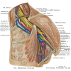
Lateral view of thorax with muscles |
 |
Nadja Baltensweiler |
Creative Commons Attribution-NonCommercial-ShareAlike |
1.976 |
1 |
|
Learning Path |
Quiz Inwendige Genitaliën Vrouw (basis) |
|
tjscherphof |
Creative Commons Attribution-NonCommercial-ShareAlike |
1.974 |
0 |
|
Learning Path |
Quiz Autonome zenuwstelsel 1 |
|
opgobee |
Creative Commons Attribution-NonCommercial-ShareAlike |
1.974 |
0 |
|
Data item |
Slagter - Drawing Cross-section of the eye - no labels |
 |
rva |
Creative Commons Attribution-NonCommercial-ShareAlike |
1.973 |
3 |
|
Data item |
Anatomy Standard - Drawing Occipital bone: anterior/lateral view - Latin labels |
 |
rva |
Creative Commons Attribution-NonCommercial |
1.972 |
1 |