
nid: 63083
Additional formats:
None available
Description:
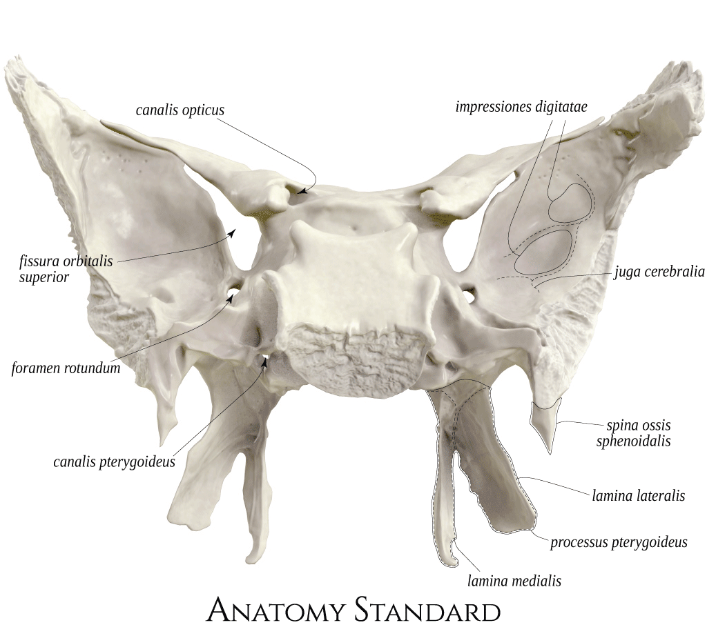
Sphenoid bone: posterior view. The sphenoid bone (os sphenoidale) is a central, unpaired bone with direct contact to almost all other skull bones. The sphenoid bone comprises the central part (corpus), the pairs of greater and lesser wings, and pterygoid processes. The dorsal aspect of the sphenoid demonstrates multiple openings and channels within the bone. The canalis opticus and fissura orbitalis superior connect the fossa cranii media with the orbit. Foramen rotundum and canalis pterygoides are the pathways to the fossa pterygopalatina. Latin labels.
Image and description retrieved from Anatomy Standard. Via this link more images can be found, including oblique views.
Image and description retrieved from Anatomy Standard. Via this link more images can be found, including oblique views.
Anatomical structures in item:
Uploaded by: rva
Netherlands, Leiden – Leiden University Medical Center, Leiden University
Os sphenoidale
Corpus ossis sphenoidalis
Ala minor ossis sphenoidalis
Ala major ossis sphenoidalis
Canalis opticus
Fissura orbitalis superior
Foramen rotundum
Canalis pterygoideus
Lamina medialis processi pterygoideus ossis sphenoidalis
Processus pterygoideus ossis sphenoidalis
Lamina lateralis processi pterygoideus ossis sphenoidalis
Spina ossis sphenoidalis
Impressiones gyrorum
Impressiones gyrorum
Creator(s)/credit: Jānis Šavlovskis MD, PhD, Assistant Professor; Kristaps Raits, 3D generalist
Requirements for usage
You are free to use this item if you follow the requirements of the license:  View license
View license If you use this item you should credit it as follows:
- For usage in print - copy and paste the line below:
- For digital usage (e.g. in PowerPoint, Impress, Word, Writer) - copy and paste the line below (optionally add the license icon):
"Anatomy Standard - Drawing Sphenoid bone: posterior view - Latin labels" at AnatomyTOOL.org by Jānis Šavlovskis and Kristaps Raits, license: Creative Commons Attribution-NonCommercial


Comments