
nid: 62371
Additional formats:
None available
Description:
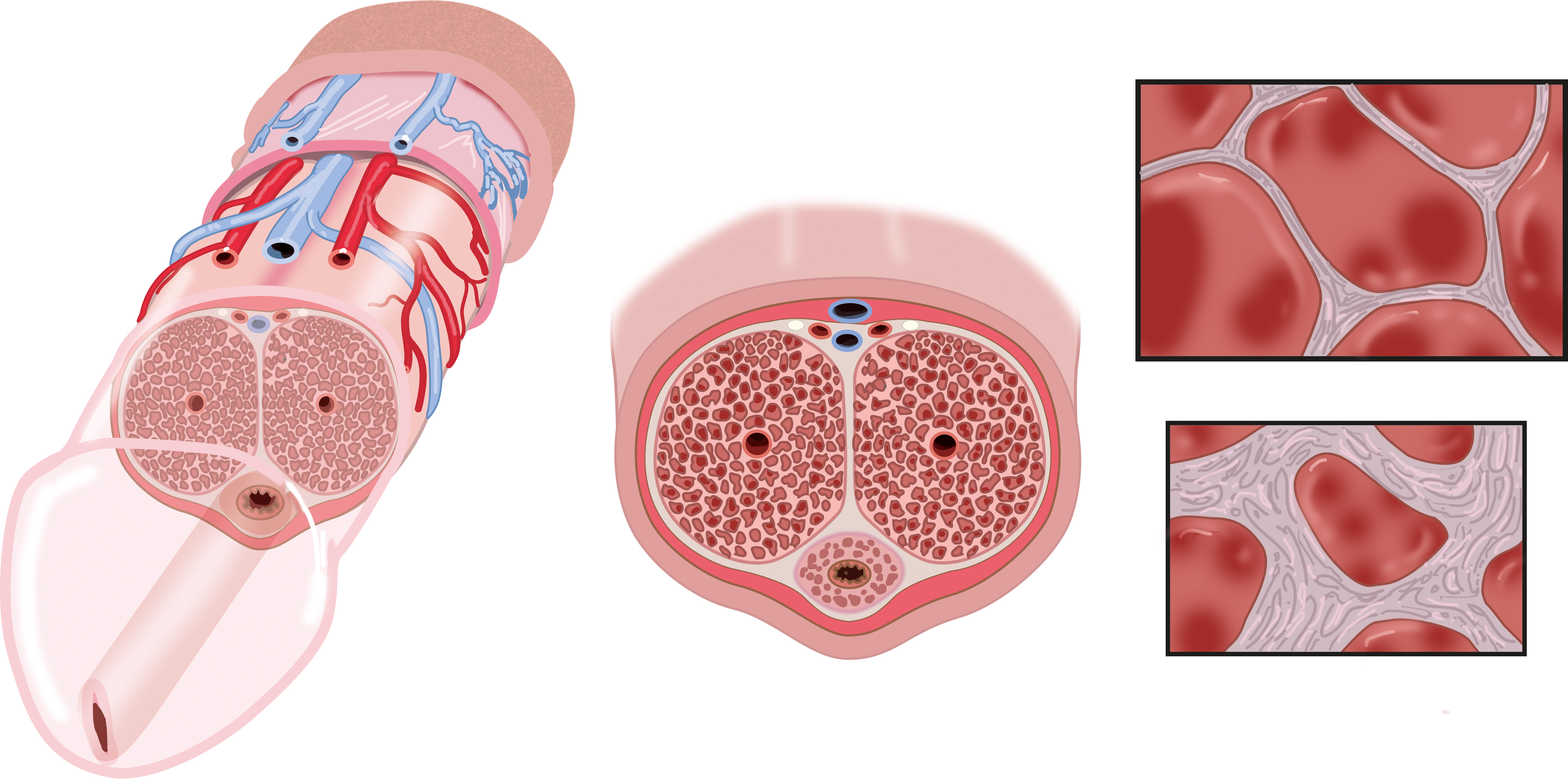
Anatomy of the penis and corpus cavernosum. The left image shows a cross-section of the penis, with the different layers and vessels on the dorsal side; the middle drawing shows a cross-section of the penis; the two right drawings show the anatomy of the corpus cavernosum, in both an erect and a non-erect situation.
Anatomical structures in item:
Uploaded by: rva
Netherlands, Leiden – Leiden University Medical Center, Leiden University
Penis
Arteria dorsalis pedis
Venae dorsales superficiales penis
Vena posterior corporis callosi
Tela subcutanea penis
Nervus dorsalis penis
Tunica albuginea corporum cavernosum (Penis)
Corpus cavernosum penis
Fascia penis
Tela subcutanea penis
Corpus spongiosum penis
Arteria profunda penis
Urethra masculina
Vena dorsalis profunda penis
Creator(s)/credit: Ron Slagter NZIMBI, medical illustrator; Prof. Marco C DeRuiter PhD, anatomist, professor of clinical and applied anatomy, LUMC
Requirements for usage
You are free to use this item if you follow the requirements of the license:  View license
View license If you use this item you should credit it as follows:
- For usage in print - copy and paste the line below:
- For digital usage (e.g. in PowerPoint, Impress, Word, Writer) - copy and paste the line below (optionally add the license icon):
"Slagter - Drawing Anatomy of the penis and corpus cavernosum - no labels" at AnatomyTOOL.org by Ron Slagter and Marco C DeRuiter, LUMC, license: Creative Commons Attribution-NonCommercial-ShareAlike


Comments