The structures in the abdomen can lie in one of three locations in relation to the peritoneum: intraperitoneal, secondary retroperitoneal or (primary) retroperitoneal. This page discusses the secondary retroperitoneal location.
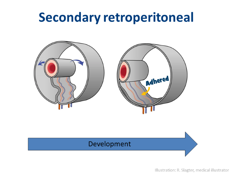
Secondary retroperitoneal structures originally lied intraperitoneally, but have been pushed aside and adhered to the body wall.
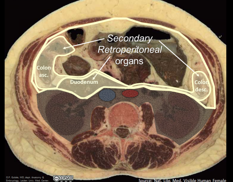
The locations of secondary retroperitoneal structures on a cross-section.
Some organs originally lied intraperitoneally, but have been pushed to the side during the embryological development. These organs and their mesenteries adhered to the abdominal wall. Upon opening the peritoneal cavity one sees them lying apparently covered by the peritoneal back wall; apparently retroperitoneally (see discussion under 'Read more' below). As they started out intraperitoneal, one calls this position: secondary retroperitoneal.
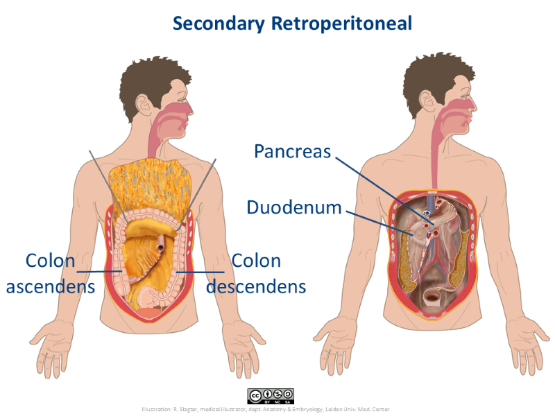
The ascending and descending colon and the duodenum and pancreas are secondary retroperitoneal organs. The left image shows the ascending and descending colon. On the right image, the colon and the stomach have been removed, to display the duodenum and pancreas.
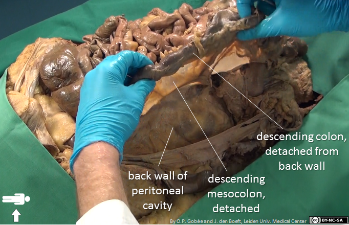
Dissection specimen. Upon opening the abdomen, one will find the descending colon and mesocolon lying adhered to the back wall of the peritoneal cavity (see the below video for that). However, they can be easily detached from the back wall again, as has been done in the specimen shown in this picture.
The ascending and descending colon and the duodenum and pancreas are secondary retroperitoneal organs. The ascending and descending colon are directly visible after one opens the peritoneal cavity, but they are fixed to the back wall, thus they are not mobile. The duodenum and pancreas lie largely hidden behind the transverse colon and the stomach. They also are adhered to the back wall.
Due to the fact that the secondary retroperitoneal organs in the very early embryological phase were still mobile and only got adhered to the back wall, they can again easily be detached from the back wall. This is a frequently applied procedure during many abdominal operations.
Secondary retroperitoneal structures in reality
In this video you see the characteristics of secondary retroperitoneal structures in a dissection specimen.
This is a fragment of a longer video that shows all three peritoneal locations. See minutes 1:56 - 2:46
(0m50s) 
Read more...
Why is it named 'retroperitoneal'?
Traditionally anatomically, it is regarded that when the mesocolon and colon parts adhere to the back wall, the mesocolon's and colon's back layer of peritoneum and the parietal peritoneum that covers the retroperitoneal space 'dissolve' and are replaced by connective tissue. Thus, only the mesocolon's and colon's front layer of peritoneum remain. The colon now lies behind this peritoneum, hence: retroperitoneal. And in this view the mesocolon is absorbed in the retroperitoneal space and disappears.
Based on the surgical observation that the secondary retroperitoneal structures can be separated from the back wall again, this traditional view is questioned. At the place of the assumedly disappeared peritoneum layers (on the back wall of the mesocolon and at the front side of the retroperitoneum), layers still are present in the adult. (Culligan 2014). Views differ as to whether those layers are still mesothelium (the characteristic covering tissue of peritoneum). If so, this would imply that the mesocolon is still present in the adult and the colon and mesocolon are not really merged into the retroperitoneal space.
Culligan K, et al. 2014. The Mesocolon. A Histological and Electron Microscopic Characterization of the Mesenteric
Attachment of the Colon Prior to and After Surgical Mobilization. Ann Surg 2014;260:1048-1056

 View license

Comments