The structures in the abdomen can lie in one of three locations in relation to the peritoneum: intraperitoneal, secondary retroperitoneal or (primary) retroperitoneal. This page discusses the extraperitoneal location, that includes the retroperitoneal location.
Extraperitoneal structures are outside the peritoneal cavity. They have been lying outside the peritoneal cavity from the very beginning of the embryological development. They are embedded in connective tissue and are therefore immobile.
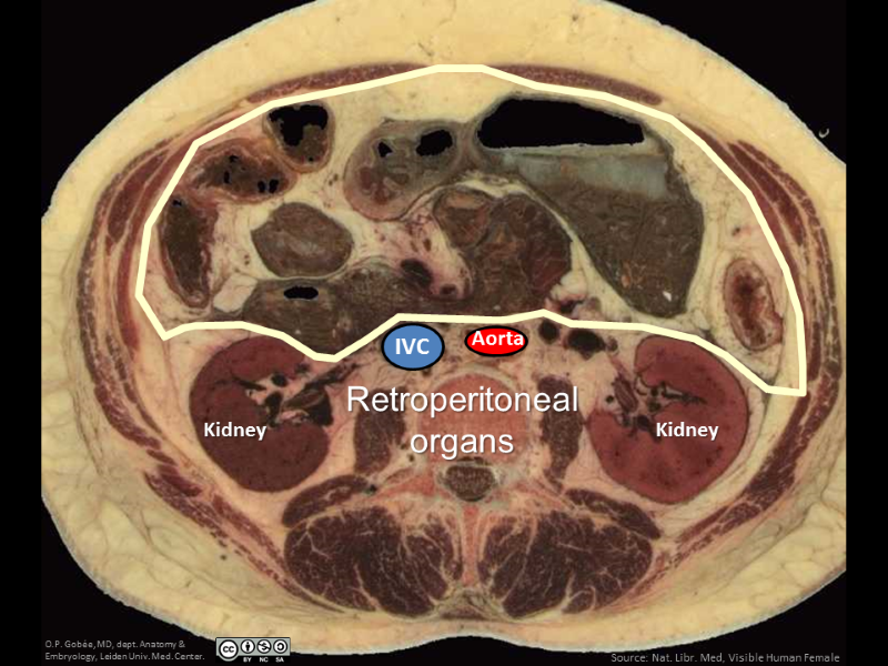
The locations of retroperitoneal structures on a cross-section. The vertebrae, aorta and inferior vena cava (IVC), and kidneys lie posterior to the peritoneum: they lie retroperitoneally.
Extraperitoneal structures lie outside the peritoneal cavity. They have been lying outside the peritoneal cavity from the very beginning of the embryological development. They are embedded in connective tissue and are therefore immobile. Moreover, as they lie outside the peritoneal cavity, one does not see them when one opens the peritoneal cavity.
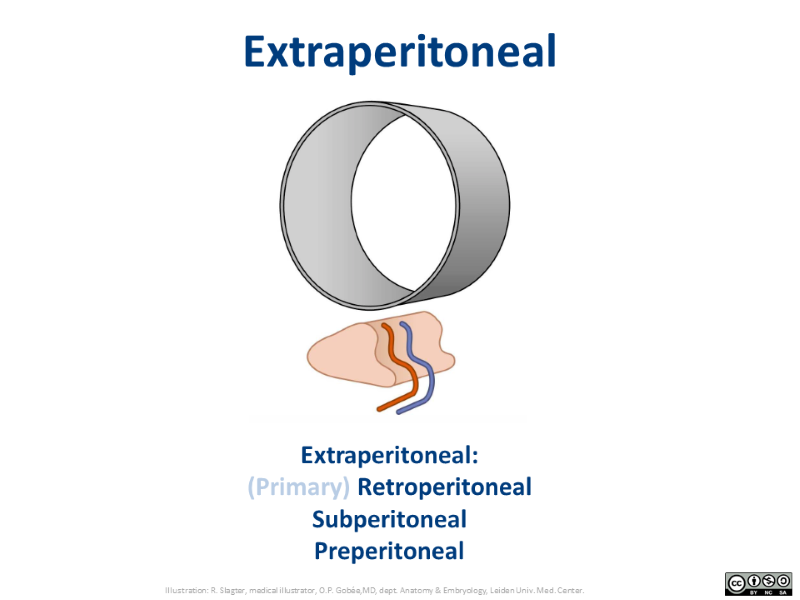
Extraperitoneal is the generic concept that comprises the more commonly used terms for the specific locations: retroperitoneal (posterior to the peritonal cavity), subperitoneal (inferior to the peritonal cavity) and preperitoneal (anterior to the peritonal cavity).
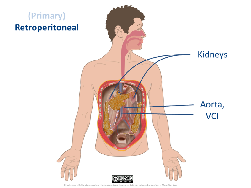
The kidneys, and the large vessels - the aorta and the inferior vena cava- are the main (primary) retroperitoneal organs.
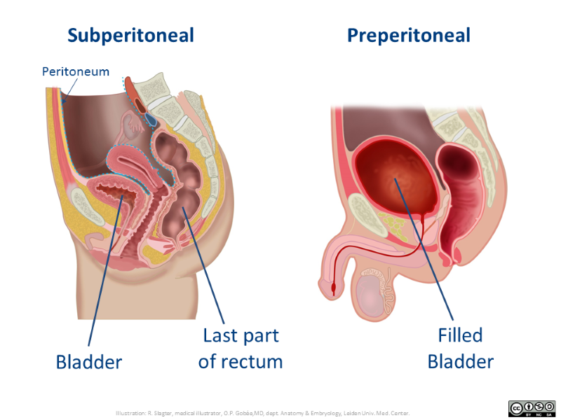
In the left image, the dashed blue line indicates the peritoneum. The bladder, the cervix of the uterus and the last part of the rectum lie subperitoneal.
The body of the uterus is surrounded by peritoneum, hence it lies intraperitoneal. The first part of the rectum lies posterior to the peritoneum, hence it is retroperitoneal.
The right image shows that the bladder, upon distention, extends between the abdominal wall and the peritoneum, thus coming to lie preperitoneal.
The kidneys, and the large vessels - the aorta and the inferior vena cava- lie posterior to the peritoneal cavity: retroperitoneal. Several pelvic organs lie inferior to the peritoneum: subperitoneal.
To access extraperitoneal organs, the surgeon can either take a route remaining completely outside or the peritoneal cavity, or traverse the peritoneal cavity, that is: cut the front wall parietal peritoneum to enter the peritoneal cavity and then cut the back wall, or inferior wall parietal peritoneum to reach respectively retroperitoneal or subperitoneal locations.
(Primary) retroperitoneal structures in reality
In this video you see the location of (primary) retroperitoneal structures in a dissection specimen. You will notice that they are not visible and not directly accessible, after opening the peritoneal cavity.
This is a fragment of a longer video that shows all three peritoneal locations. See minutes 2:46 - 4:00
(1m14s) 

 View license

Comments