|
Data item |
LadyofHats - Drawing Overview of the gastrointestinal tract - No labels |
 |
rva |
Public Domain |
760 |
2 |
|
Data item |
Leiden - Student - Video Anatomy of the heart - Dutch spoken, Latin labels |
|
Flynn Post |
Creative Commons Attribution-ShareAlike |
759 |
1 |
|
Data item |
Radiopaedia - Drawing Innervation (supra)scapular - English labels |
 |
rva |
Creative Commons Attribution-NonCommercial-NoDerivs |
759 |
1 |
|
Anatomical Structure |
Labyrinthus ethmoidalis |
|
admin |
|
759 |
1 |
|
Data item |
3D Anatomy Lyon: The hyoid bone and cartilages of the larynx - video of 3D model |
|
rva |
Creative Commons Attribution-NonCommercial-NoDerivs |
759 |
2 |
|
Data item |
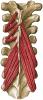
Sobotta 1909 fig.240 - transverso-spinal muscles - no labels |
 |
rva |
Creative Commons Attribution-ShareAlike |
758 |
2 |
|
Data item |
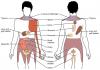
Slagter - Drawing Impingement of shoulder - no labels |
 |
rva |
Creative Commons Attribution-NonCommercial-ShareAlike |
758 |
1 |
|
Anatomical Structure |
Vena mediana cubiti |
|
admin |
|
758 |
1 |
|
Learning Path |
Quiz LUMC OA-AM M1 Hart - Kenmerken en onderdelen |
|
opgobee |
Creative Commons Attribution-NonCommercial-ShareAlike |
758 |
0 |
|
Data item |
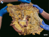
Leiden- Drawing Spleen with splenorenal and gastrosplenic ligaments - no labels |
 |
opgobee |
Creative Commons Attribution-NonCommercial-ShareAlike |
758 |
2 |
|
Data item |
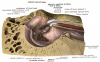
U.Br.Columbia - Photo Deep gluteal region of the lower limb (dissection) |
|
rva |
Creative Commons Attribution-NonCommercial-ShareAlike |
757 |
3 |
|
Anatomical Structure |
Vena cava superior |
|
admin |
|
757 |
2 |
|
Data item |
OpenStax AnatPhys fig.15.7 - Referred Pain Chart - English labels |
 |
Jorn IJkhout |
Creative Commons Attribution |
757 |
3 |
|
Anatomical Structure |
Palpebra inferior |
|
admin |
|
757 |
1 |
|
Data item |
3D Anatomy Lyon: Proprioception of muscles, tendons and joints - video of 3D model |
|
rva |
Creative Commons Attribution-NonCommercial-NoDerivs |
757 |
2 |
|
Anatomical Structure |
Meatus nasi communis |
|
admin |
|
757 |
3 |
|
Data item |
Gent - Video Anatomie van het colon (dissectie-preparaat) |
|
rva |
Creative Commons Attribution-NonCommercial-ShareAlike |
757 |
2 |
|
Data item |
Radiopaedia - Drawing The spring (plantar calcaneonavicular) ligament complex - English labels |
 |
rva |
Creative Commons Attribution-NonCommercial-NoDerivs |
757 |
1 |
|
Anatomical Structure |
Sulcus intertubercularis |
|
admin |
|
756 |
2 |
|
Data item |
Sobotta 1914 fig.823 - The female breast, anterior view - English labels |
 |
Student128 |
Public Domain |
756 |
2 |
|
Data item |
Servier - Drawing Thyroid gland - no labels |
 |
rva |
Creative Commons Attribution |
756 |
1 |
|
Data item |
Radiopaedia - Drawing Parietal and visceral pleura of lung - English labels |
 |
rva |
Creative Commons Attribution-NonCommercial-NoDerivs |
755 |
5 |
|
Anatomical Structure |
Cranium |
|
admin |
|
755 |
1 |
|
Data item |
Alveolus |
 |
opgobee |
Public Domain |
755 |
3 |
|
Anatomical Structure |
Pars petrosa (Arteria carotis interna) |
|
admin |
|
755 |
2 |
|
Data item |
Leiden - Photo Colon, mesocolon, marginal artery (dissection) - Latin labels |
 |
opgobee |
Creative Commons Attribution-NonCommercial-ShareAlike |
754 |
1 |
|
Data item |
Sobotta 1909 fig.714 - Nerves and vessels of the nuchal region, superficial and middle layer - English labels |
 |
Student128 |
Public Domain |
754 |
2 |
|
Anatomical Structure |
Basis cranii externa |
|
admin |
|
754 |
1 |
|
Anatomical Structure |
Concha nasalis suprema |
|
admin |
|
754 |
2 |
|
Anatomical Structure |
Incisura ethmoidalis |
|
admin |
|
754 |
1 |
|
Data item |
Sobotta 1909 fig.192 - sternoclavicular joints and costosternal articulations, anterior view - English Labels |
 |
Student128 |
Public Domain |
753 |
4 |
|
Data item |
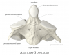
Anatomy Standard - Drawing Axis: anterior aspect - Latin labels |
 |
rva |
Creative Commons Attribution-NonCommercial |
753 |
2 |
|
Anatomical Structure |
Linea mylohyoidea |
|
admin |
|
753 |
1 |
|
Data item |
3D Anatomy Lyon: The quadriceps muscle - video of 3D model |
|
rva |
Creative Commons Attribution-NonCommercial-NoDerivs |
753 |
14 |
|
Anatomical Structure |
Processus uncinatus pancreatis |
|
admin |
|
753 |
2 |
|
Anatomical Structure |
Plicae circulares intestini tenuis |
|
admin |
|
752 |
1 |
|
Anatomical Structure |
Calvaria |
|
admin |
|
752 |
1 |
|
Data item |
Cenveo - Drawing Location of salivary glands and ducts - English labels |
 |
rva |
Creative Commons Attribution |
751 |
3 |
|
Data item |
Palmer - Drawing Muscles and triangles of neck - English labels |
 |
rva |
Creative Commons Attribution |
751 |
1 |
|
Anatomical Structure |
Auris interna |
|
admin |
|
751 |
0 |
|
Data item |
3D Anatomy Lyon: Arthrology of the pelvis in childbirth - video of 3D model |
|
rva |
Creative Commons Attribution-NonCommercial-NoDerivs |
751 |
2 |
|
Data item |
Radiopaedia - Drawing Bones of the knee joint - no labels |
 |
rva |
Creative Commons Attribution-NonCommercial-ShareAlike |
751 |
2 |
|
Anatomical Structure |
Arteria choroidea anterior |
|
admin |
|
751 |
1 |
|
Data item |
Sobotta 1909 fig.713 - Superficial nerves and vessels of the back of the hand - English labels |
 |
Student128 |
Public Domain |
751 |
2 |
|
Data item |
Slagter - Drawing Sagittal section of the eye and muscles of eye - no labels |
 |
rva |
Creative Commons Attribution-NonCommercial-ShareAlike |
751 |
1 |
|
Data item |
Newey - 3D model Kidney with Polycystic Kidney Disease (ADPKD) compared to a healthy kidney |
|
rva |
Creative Commons Attribution-NonCommercial-NoDerivs |
750 |
3 |
|
Data item |
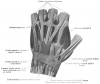
Sobotta 1909 fig.289 - Tendons and tendon-sheats of the extensor muscles in the hand - English labels |
 |
Student128 |
Public Domain |
750 |
1 |
|
Data item |
Anatomy Standard - Drawing Nasal cavity: sagittal cut - Latin labels |
 |
rva |
Creative Commons Attribution-NonCommercial |
750 |
1 |
|
Anatomical Structure |
Pars prostatica |
|
admin |
|
750 |
1 |
|
Anatomical Structure |
Sella turcica |
|
admin |
|
749 |
1 |
|
Anatomical Structure |
Sutura parietomastoidea |
|
admin |
|
749 |
1 |
|
Data item |
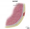
Slagter- Drawing Pneumothorax mechanism - no labels |
 |
kmeier |
Creative Commons Attribution-NonCommercial-ShareAlike |
749 |
1 |
|
Anatomical Structure |
Ductus biliaris |
|
admin |
|
749 |
0 |
|
Anatomical Structure |
Margo posterior partis petrosae ossis temporalis |
|
admin |
|
748 |
0 |
|
Anatomical Structure |
Capsula adiposa perirenalis |
|
admin |
|
748 |
0 |
|
Data item |
Servier - Drawing Suprachiasmatic nucleus of hypothalamus - no labels |
 |
rva |
Creative Commons Attribution |
748 |
2 |
|
Data item |
Anatomy Standard - Drawing Lateral wall of nasal cavity: sagittal cut - Latin labels |
 |
rva |
Creative Commons Attribution-NonCommercial |
748 |
1 |
|
Anatomical Structure |
Canalis caroticus |
|
admin |
|
747 |
1 |
|
Anatomical Structure |
Sutura cranii |
|
admin |
|
747 |
2 |
|
Anatomical Structure |
Ramus dexter vena portae hepatis |
|
admin |
|
747 |
1 |
|
Data item |
Sobotta 1909 fig.244 - musculi intertransversarii - English labels |
 |
rva |
Creative Commons Attribution-ShareAlike |
747 |
1 |
|
Anatomical Structure |
Facies articularis posterior dentis |
|
admin |
|
747 |
1 |
|
Anatomical Structure |
Sinus durae matris |
|
admin |
|
747 |
2 |
|
Anatomical Structure |
Musculus epicranius |
|
admin |
|
746 |
0 |
|
Data item |
arteria mesenterica superior |
 |
aherrler |
Creative Commons Attribution-ShareAlike |
746 |
2 |
|
Data item |
Leiden - Photo Colon, mesocolon, marginal artery (dissection) - no labels |
 - no labels") |
opgobee |
Creative Commons Attribution-NonCommercial-ShareAlike |
746 |
1 |
|
Data item |
Leiden, Maas Photo 2 - Sagittal section of the female pelvis (plastination specimen) - number labels |
 |
rva |
Creative Commons Attribution-NonCommercial-ShareAlike |
746 |
1 |
|
Data item |
Cenveo - Drawing Structure of the photoreceptor cells - English labels |
 |
rva |
Creative Commons Attribution |
745 |
3 |
|
Anatomical Structure |
Arteria iliaca interna |
|
admin |
|
745 |
2 |
|
Data item |
Universiteit Gent - Students - Photo Gaster/Stomach (4) - numbered Latin labels |
 |
sehellin |
Creative Commons Attribution-NonCommercial-ShareAlike |
745 |
1 |
|
Anatomical Structure |
Circumferentia articularis capitis ulnae |
|
admin |
|
745 |
0 |
|
Anatomical Structure |
Splen |
|
admin |
|
745 |
1 |
|
Data item |
Anatomy Standard - Drawing Anterior aspect of cervical vertebra (C3) - Latin labels |
 |
rva |
Creative Commons Attribution-NonCommercial |
745 |
1 |
|
Data item |
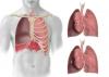
OpenStax AnatPhys fig.22.14 - The Lung Pleurea - English labels |
 |
Jorn IJkhout |
Creative Commons Attribution |
745 |
1 |
|
Data item |
Leiden - Drawing Heart: anterior view and cross-section - Dutch labels |
 |
rva |
Creative Commons Attribution-NonCommercial-ShareAlike |
745 |
1 |
|
Data item |
Sobotta 1911 fig.804 - Inner wall of the right tympanic cavity, lateral view - English labels |
 |
Student128 |
Public Domain |
744 |
3 |
|
Data item |
Radiopaedia - Drawing Boundaries of the femoral triangle - English labels |
 |
rva |
Creative Commons Attribution-NonCommercial-ShareAlike |
744 |
2 |
|
Data item |
OpenStax AnatPhys fig.27.12 - Folliculogenesis - English labels |
 |
Jorn IJkhout |
Creative Commons Attribution |
744 |
3 |
|
Anatomical Structure |
Valvulae anales |
|
admin |
|
744 |
2 |
|
Anatomical Structure |
Septum nasi osseum |
|
admin |
|
744 |
1 |
|
Data item |
Groningen - 3D model Thorax and abdomen, a few important muscles |
|
rva |
Creative Commons Attribution-NonCommercial-ShareAlike |
744 |
2 |
|
Data item |
Blausen - Forebrain, midbrain, hindbrain - English labels |
 |
Student10 |
Creative Commons Attribution-ShareAlike |
744 |
3 |
|
Anatomical Structure |
Linea pectinata canalis analis |
|
admin |
|
744 |
1 |
|
Data item |
Internal View on the Female Endopelvic Fascia - no labels |
 |
opgobee |
Creative Commons Attribution-NonCommercial-ShareAlike |
743 |
4 |
|
Data item |
Slagter - Drawing Pelvis and hip joint - no labels |
 |
rva |
Creative Commons Attribution-NonCommercial-ShareAlike |
743 |
3 |
|
Data item |
Radiopaedia - Drawing Rectus sheath below arcuate line - no labels |
 |
rva |
Creative Commons Attribution-NonCommercial-ShareAlike |
743 |
3 |
|
Data item |
Anatomy Standard - Drawing Sphenoid bone: posterior view - no labels |
 |
rva |
Creative Commons Attribution-NonCommercial |
743 |
5 |
|
Anatomical Structure |
Fossa sacci lacrimalis |
|
admin |
|
743 |
1 |
|
Data item |
U.Br.Columbia - Drawing Cross section of cerebellum: middle cerebellar peduncle - English labels |
 |
rva |
Creative Commons Attribution-NonCommercial-ShareAlike |
742 |
1 |
|
Anatomical Structure |
Flexura sacralis recti |
|
admin |
|
742 |
2 |
|
Data item |
Blausen 0732 - Female internal genital organs - English labels |
 |
Student10 |
Creative Commons Attribution |
741 |
1 |
|
Anatomical Structure |
Camera vitrea bulbi oculi |
|
admin |
|
741 |
1 |
|
Data item |
Anatomy Standard - Drawing First metatarsal bone - Latin labels |
 |
rva |
Creative Commons Attribution-NonCommercial |
741 |
5 |
|
Anatomical Structure |
Scapula |
|
admin |
|
741 |
1 |
|
Anatomical Structure |
Sulcus arteriae temporalis mediae |
|
admin |
|
740 |
1 |
|
Anatomical Structure |
Musculus pterygoideus lateralis |
|
admin |
|
740 |
0 |
|
Anatomical Structure |
Norma inferior cranii |
|
admin |
|
739 |
1 |
|
Anatomical Structure |
Crista occipitalis externa |
|
admin |
|
739 |
1 |
|
Anatomical Structure |
Tunica fibrosa bulbi |
|
admin |
|
739 |
2 |
|
Data item |
Sobotta 1909 fig.696 - Nerves and vessels of the nuchal region, deep layer - English labels |
 |
Student128 |
Public Domain |
739 |
1 |