|
Data item |
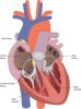
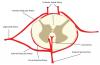
Slagter - Drawing Position of sinoatrial and atrioventricular node - Dutch labels |
 |
rva |
Creative Commons Attribution-NonCommercial-ShareAlike |
644 |
4 |
|
Data item |
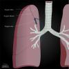
Radiopaedia - Drawing Azygos lobe - English labels |
 |
rva |
Creative Commons Attribution-NonCommercial-ShareAlike |
644 |
4 |
|
Data item |
Leiden - Video The Phrenicooesophageal ligament |
|
opgobee |
Creative Commons Attribution-NonCommercial-ShareAlike |
644 |
2 |
|
Data item |
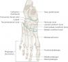
Palmer - Drawing Bones and joints of the foot - English labels |
 |
rva |
Creative Commons Attribution |
644 |
1 |
|
Anatomical Structure |
Processus uncinatus vertebrae cervicales |
|
admin |
|
644 |
1 |
|
Anatomical Structure |
Vena epigastrica inferior |
|
admin |
|
644 |
1 |
|
Anatomical Structure |
Cortex renalis |
|
admin |
|
644 |
3 |
|
Data item |
Slagter - Drawing Peritoneal reflections on the female internal pelvic organs - No labels |
 |
admin |
Creative Commons Attribution-NonCommercial-ShareAlike |
644 |
1 |
|
Data item |
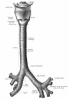
Sobotta 1906 fig.443 - Larynx, trachea and bronchi, anterior view - English labels |
 |
Student128 |
Public Domain |
644 |
3 |
|
Data item |
3D Anatomy Lyon: Sacrum and coccyx: anatomy and nerve paths - video of 3D model |
|
rva |
Creative Commons Attribution-NonCommercial-NoDerivs |
643 |
3 |
|
Data item |
artificial 3D Head with mimic muscles and facial nerve |
|
aherrler |
Creative Commons Attribution-NonCommercial-ShareAlike |
643 |
1 |
|
Data item |
Radiopaedia - Drawing Scapula superior view - English labels |
 |
rva |
Creative Commons Attribution-NonCommercial-NoDerivs |
643 |
3 |
|
Data item |
RCSI - Drawing Arteries and veins of kidney and adrenal gland - no labels |
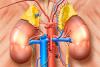 |
rva |
Creative Commons Attribution-NonCommercial-ShareAlike |
643 |
2 |
|
Anatomical Structure |
Fissura tympanomastoidea |
|
admin |
|
643 |
0 |
|
Anatomical Structure |
Ductus biliferi interlobulares |
|
admin |
|
643 |
2 |
|
Anatomical Structure |
Ostium urethrae internum (Urethra masculina) |
|
admin |
|
643 |
2 |
|
Anatomical Structure |
Arteria transversa cervicis |
|
admin |
|
643 |
0 |
|
Anatomical Structure |
Arteria gastroomentalis dextra |
|
admin |
|
643 |
1 |
|
Anatomical Structure |
Ramus carpalis dorsalis ateria radialis |
|
admin |
|
642 |
1 |
|
Data item |
Anatomy Standard - Drawing Bones of foot: dorsal view - Latin labels |
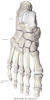 |
rva |
Creative Commons Attribution-NonCommercial |
642 |
3 |
|
Anatomical Structure |
Costae verae [I-VII] |
|
admin |
|
642 |
0 |
|
Learning Path |
Quiz LUMC HC Abdomen Gobée |
|
opgobee |
Creative Commons Attribution-NonCommercial-ShareAlike |
642 |
1 |
|
Anatomical Structure |
Ligamentum falciforme hepatis |
|
admin |
|
642 |
0 |
|
Data item |
Radiopaedia - Drawing Muscle borders of the femoral triangle - English labels |
 |
rva |
Creative Commons Attribution-NonCommercial-ShareAlike |
641 |
1 |
|
Data item |
Radiopaedia - Drawing Deltoid ligament of ankle: superficial layer - English labels |
 |
rva |
Creative Commons Attribution-NonCommercial-NoDerivs |
641 |
2 |
|
Data item |
Anatomy Standard - Drawing Apertura thoracis superior (thoracic inlet) - no labels |
 |
rva |
Creative Commons Attribution-NonCommercial |
641 |
4 |
|
Anatomical Structure |
Fascia renalis |
|
admin |
|
641 |
0 |
|
Data item |
Sobotta 1909 fig.90 - mandible, anterior view - English Labels |
 |
Student128 |
Public Domain |
641 |
3 |
|
Data item |
3D Anatomy Lyon: The sternum - video of 3D model |
|
rva |
Creative Commons Attribution-NonCommercial-NoDerivs |
641 |
2 |
|
Data item |
KnowledgeWorks - Drawing Location of the parathyroid glands - English labels |
 |
rva |
Creative Commons Attribution |
640 |
1 |
|
Data item |
U.Br.Columbia - Drawing Lateral view of brain - English labels |
 |
rva |
Creative Commons Attribution-NonCommercial-ShareAlike |
640 |
4 |
|
Data item |
Slagter - Drawing Endochondral ossification (bone formation) - no labels |
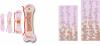 |
rva |
Creative Commons Attribution-NonCommercial-ShareAlike |
640 |
3 |
|
Data item |
Sobotta 1909 fig.684 - Ramification of the trigeminal nerve with communications, schematic - English labels |
 |
Student128 |
Public Domain |
640 |
2 |
|
Anatomical Structure |
Cervix uteri |
|
admin |
|
640 |
1 |
|
Anatomical Structure |
Facies articularis fossae mandibularis ossis temporalis |
|
admin |
|
640 |
1 |
|
Data item |
Sobotta 1906 fig.414 - Decending colon and sigmoid with mesocolon - English labels |
 |
Student128 |
Public Domain |
640 |
5 |
|
Anatomical Structure |
Margo medialis renis |
|
admin |
|
640 |
0 |
|
Anatomical Structure |
Sulcus nervi ulnaris |
|
admin |
|
640 |
1 |
|
Anatomical Structure |
Vena iliaca externa |
|
admin |
|
640 |
2 |
|
Data item |
testis mouse |
|
aherrler |
Creative Commons Attribution-NonCommercial-ShareAlike |
640 |
2 |
|
Data item |
MedicalGraphics - Drawing Segments of the kidney - no labels |
 |
rva |
Creative Commons Attribution-NoDerivatives |
639 |
3 |
|
Data item |
MedicalGraphics - Drawing Bones of arm: anterior view - no labels |
 |
rva |
Creative Commons Attribution-NoDerivatives |
639 |
2 |
|
Data item |
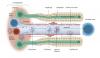
Cenveo - Drawing Depiction of the visual system of the central nervous system - English labels |
 |
rva |
Creative Commons Attribution |
639 |
1 |
|
Data item |
Anatomy Standard - Drawing Sternum: frontal and anterolateral view - no labels |
 |
rva |
Creative Commons Attribution-NonCommercial |
639 |
7 |
|
Data item |
Sobotta 1906 fig.442 - Muscles of the larynx, lateral view - English labels |
 |
Student128 |
Public Domain |
639 |
3 |
|
Data item |
Sobotta 1909 fig.271 - deep muscles of the arm, lateral view - English Labels |
 |
Student128 |
Public Domain |
639 |
1 |
|
Data item |
U.Br.Columbia - Module Brachial plexus |
|
rva |
Creative Commons Attribution-NonCommercial-ShareAlike |
639 |
1 |
|
Data item |
Sobotta 1909 fig.603 - Lymph nodes of the head and neck of a child - English labels |
 |
Student128 |
Public Domain |
638 |
3 |
|
Anatomical Structure |
Sulcus hamuli pterygoidei |
|
admin |
|
638 |
1 |
|
Anatomical Structure |
Arteria phrenica inferior |
|
admin |
|
638 |
2 |
|
Data item |
Blausen 0393 - Facial bones - English labels |
 |
Student10 |
Creative Commons Attribution |
638 |
2 |
|
Data item |
Anatomy Standard - Drawing Costotransversal joints with ligament and capsule - Latin labels |
 |
rva |
Creative Commons Attribution-NonCommercial |
638 |
2 |
|
Anatomical Structure |
Musculi auriculares |
|
admin |
|
638 |
4 |
|
Data item |
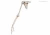
Slagter - Drawing Bones upper limb dorsal side - no labels |
 |
lumcanatomy |
Creative Commons Attribution-NonCommercial-ShareAlike |
638 |
4 |
|
Data item |
Radiopaedia - Drawing Cross-section of the penis - no labels |
 |
rva |
Creative Commons Attribution-NonCommercial-ShareAlike |
638 |
3 |
|
Anatomical Structure |
Vomer |
|
admin |
|
638 |
2 |
|
Data item |
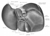
Sobotta 1906 fig.397 - Spleen, hilus surface - English labels |
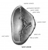 |
Student128 |
Public Domain |
638 |
1 |
|
Anatomical Structure |
Columnae anales |
|
admin |
|
638 |
2 |
|
Data item |
Anatomy Standard - Drawing Axis: anterior aspect - no labels |
 |
rva |
Creative Commons Attribution-NonCommercial |
637 |
3 |
|
Anatomical Structure |
Fascia penis |
|
admin |
|
637 |
2 |
|
Anatomical Structure |
Ductus hepaticus sinister |
|
admin |
|
637 |
0 |
|
Anatomical Structure |
Tunica serosa (Vesica urinaria) |
|
admin |
|
637 |
0 |
|
Data item |
OpenStax AnatPhys fig.6.7 - Anatomy of a Long Bone - English labels |
 |
Jorn IJkhout |
Creative Commons Attribution |
637 |
2 |
|
Anatomical Structure |
Camerae bulbi oculi |
|
admin |
|
637 |
2 |
|
Data item |
Leiden - Drawing Dorsal view of embryonal and adult heart - English labels |
 |
rva |
Creative Commons Attribution-NonCommercial-ShareAlike |
637 |
2 |
|
Data item |
StatPearls - Drawing Spinal arteries in transverse plane - English labels |
 |
rva |
Creative Commons Attribution-NonCommercial-NoDerivs |
637 |
3 |
|
Data item |
Slagter - Drawing Portal field and hepatocytes - Dutch labels |
 |
rva |
Creative Commons Attribution-NonCommercial-ShareAlike |
637 |
3 |
|
Anatomical Structure |
Canalis vomerorostralis |
|
admin |
|
637 |
1 |
|
Data item |
Universiteit Gent - Students - Drawing Bursa omentalis (1) - no labels |
 |
sehellin |
Creative Commons Attribution-NonCommercial-ShareAlike |
636 |
3 |
|
Interactive content |
(Prosection Thorax) - Visceral and parietal pleura |
|
holtder |
Creative Commons Attribution |
636 |
2 |
|
Learning Path |
SIP Practice Exam BBS1005 |
|
lilysokolova |
Creative Commons Attribution-NonCommercial-ShareAlike |
636 |
1 |
|
Data item |
Sobotta 1906 fig.389 - Liver, inferior view - English labels |
 |
Student128 |
Public Domain |
636 |
1 |
|
Anatomical Structure |
Sulcus sinus occipitalis |
|
admin |
|
636 |
1 |
|
Data item |
Sobotta 1909 fig.7 - atlas, superior view - English Labels |
 |
Student128 |
Public Domain |
636 |
3 |
|
Data item |
Sobotta 1909 fig.229 - ligaments of the foot, plantar surface - English Labels |
 |
Student128 |
Public Domain |
636 |
2 |
|
Data item |
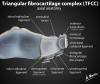
Radiopaedia - Drawing Triangular fibrocartilage complex - English labels |
 |
rva |
Creative Commons Attribution-NonCommercial-ShareAlike |
636 |
4 |
|
Data item |
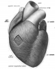
Sobotta 1906 fig.524 - musculature of the heart, anterior view - English Labels |
 |
Student128 |
Public Domain |
636 |
1 |
|
Anatomical Structure |
Canalis pudendalis |
|
admin |
|
635 |
0 |
|
Data item |
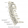
Anatomy Standard - Drawing Cervical Part of Columna Vertebralis (C1-C7): lateral aspect - Latin labels |
 |
rva |
Creative Commons Attribution-NonCommercial |
635 |
3 |
|
Data item |
Leiden - Photo Internal view Jejunum with circular folds - no labels |
 |
opgobee |
Creative Commons Attribution-NonCommercial-ShareAlike |
635 |
2 |
|
Anatomical Structure |
Crus clitoridis |
|
admin |
|
635 |
2 |
|
Interactive content |
main bronchi |
|
A.Agten |
Creative Commons Attribution-ShareAlike |
635 |
1 |
|
Data item |
Leiden - Drawing Congenital heart defects: transposition of the great arteries, ventricular septal defect (closed semilunar valves) - no labels |
 |
rva |
Creative Commons Attribution-NonCommercial-ShareAlike |
635 |
2 |
|
Data item |
Sobotta 1911 fig.725 - Nerves and vessels of the female perineum - English labels |
 |
Student128 |
Public Domain |
635 |
2 |
|
Anatomical Structure |
Fundus vesicae biliaris |
|
admin |
|
635 |
0 |
|
Data item |
Leiden-Delft-Groningen - 3D model Transposition of the great arteries after switch - numbered English labels |
|
opgobee |
Creative Commons Attribution-NonCommercial-ShareAlike |
634 |
2 |
|
Anatomical Structure |
Fascia pharyngobasilaris |
|
admin |
|
634 |
0 |
|
Anatomical Structure |
Extremitas superior (Testis) |
|
admin |
|
634 |
2 |
|
Anatomical Structure |
Arteria communicans posterior |
|
admin |
|
634 |
3 |
|
Data item |
Sobotta 1909 fig.103 - pterygopalatine fossa, lateral view - English Labels |
 |
Student128 |
Public Domain |
634 |
1 |
|
Data item |
Anatomy Standard - Drawing Right palatine bone: lateral view - Latin labels |
 |
rva |
Creative Commons Attribution-NonCommercial |
634 |
3 |
|
Anatomical Structure |
Arteria testicularis <male> |
|
admin |
|
634 |
0 |
|
Data item |
Anatomy Standard - Drawing Mandibula: anterolateral view - no labels |
 |
rva |
Creative Commons Attribution-NonCommercial |
633 |
4 |
|
Data item |
Blausen 0865 - Anatomy of the trachea - English labels |
 |
Student10 |
Creative Commons Attribution |
633 |
2 |
|
Interactive content |
brain sagittal |
|
aherrler |
Creative Commons Attribution-NonCommercial-ShareAlike |
633 |
2 |
|
Anatomical Structure |
Hilum lienale |
|
admin |
|
633 |
0 |
|
Anatomical Structure |
Tegmen tympani |
|
admin |
|
633 |
0 |
|
Data item |
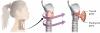
RCSI - Drawing Thyroid and parathyroid glands and vasculature: anterior and lateral - English labels |
 |
rva |
Creative Commons Attribution-NonCommercial-ShareAlike |
633 |
4 |
|
Data item |
Cenveo - Drawing Dually innervated heart and lungs - English labels |
 |
rva |
Creative Commons Attribution |
633 |
2 |
|
Data item |
Slagter - Drawing Muscles of thigh: posterior view - no labels |
 |
rva |
Creative Commons Attribution-NonCommercial-ShareAlike |
633 |
3 |