
Serous membranes
The pleura, pericardium and peritoneum are serous membranes. This section explains the terms 'serous membrane', 'serosa', 'mesothelium', which are often used in close relation with each other.
Serous membranes are membranes lining closed internal body cavities. The pleura, pericardium and peritoneum are serous membranes that line respectively the pleural, pericardial and peritoneal cavities.
Serous membranes secrete a slight amount of lubricating fluid. This allows the layers of the pleura, pericardium and peritoneum to move in relation to each other, and hence provides a certain amount of mobility to the ensheathed organs (resp. lung, heart, intestine). The secreted fluid is called serous fluid. A serous fluid is a watery fluid, resembling (blood-)serum. This also explains the name 'serous membrane'.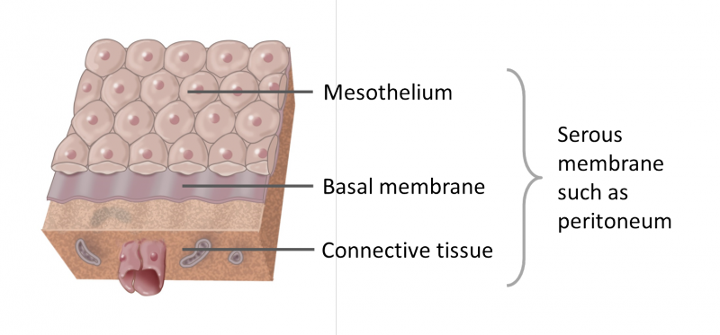
Figure. Histology of a serous membrane
Serous membranes consist of a single layer of epithelium, named mesothelium, attached to a supporting layer of connective tissue, with a small layer in between, the basal membrane (fig 1).
Epithelia are covering tissues. The type of epithelium that lines the internal body cavities, is called mesothelium. It is the mesothelium that secretes the lubricating fluid.
The largest part of the gut tube is ensheathed in peritoneum. Histologically, this can be seen as a layer on the outside of the gut. In histology this layer is called serosa after serous membrane. Serosa thus is the same as visceral peritoneum.
 View license

Comments