nid: 63011
Additional formats:
None available
Description:
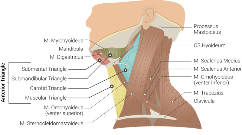
Muscles and triangles of neck. This illustration shows several muscles of the neck, as well as the different parts of the anterior triangle. English labels
Figure retrieved from AlShareef S, Newton BW. Accessory Nerve Injury. [Updated 2022 May 1]. In: StatPearls [Internet]. Treasure Island (FL): StatPearls Publishing; 2022 Jan-. Available from: https://www.ncbi.nlm.nih.gov/books/NBK532245/ (CC BY)
Figure retrieved from AlShareef S, Newton BW. Accessory Nerve Injury. [Updated 2022 May 1]. In: StatPearls [Internet]. Treasure Island (FL): StatPearls Publishing; 2022 Jan-. Available from: https://www.ncbi.nlm.nih.gov/books/NBK532245/ (CC BY)
Anatomical structures in item:
Uploaded by: rva
Netherlands, Leiden – Leiden University Medical Center, Leiden University
Collum
Musculus mylohyoideus
Mandibula
Musculus digastricus
Trigonum submentale
Trigonum cervicale anterius
Trigonum submandibulare
Trigonum caroticum
Trigonum omotracheale
Musculus omohyoideus
Venter superior musculus omohyoidei
Musculus sternocleidomastoideus
Processus mastoideus
Os hyoideum
Musculus scalenus medius
Musculus scalenus anterior
Venter superficialis musculus omohyoidei inferior
Musculus trapezius
Clavicula
Creator(s)/credit: Beckie Palmer
Requirements for usage
You are free to use this item if you follow the requirements of the license:  View license
 View license
 If you use this item you should credit it as follows:
- For usage in print - copy and paste the line below:
- For digital usage (e.g. in PowerPoint, Impress, Word, Writer) - copy and paste the line below (optionally add the license icon):
"Palmer - Drawing Muscles and triangles of neck - English labels" at AnatomyTOOL.org by Beckie Palmer, © StatPearls Publishing LLC, license: Creative Commons Attribution
"Palmer - Drawing Muscles and triangles of neck - English labels" by Beckie Palmer, © StatPearls Publishing LLC, license: CC BY


Comments