|
Data item |
Radiopaedia - Drawing Radial recurrent artery medial view - English labels |
 |
rva |
Creative Commons Attribution-NonCommercial-NoDerivs |
1.151 |
1 |
|
Anatomical Structure |
Linea axillaris anterior |
|
admin |
|
1.151 |
0 |
|
Data item |
Sobotta 1909 fig.689 - Trigeminal nerve, sphenopalatine ganglion, facial and tympanic nerves - English labels |
 |
Student128 |
Public Domain |
1.150 |
3 |
|
Data item |
OpenStax AnatPhys fig.11.17 - The Diaphragm and crura - Latin, Dutch labels |
 |
admin |
Creative Commons Attribution |
1.149 |
1 |
|
Data item |
Radiopaedia - Drawing Contents of cubital fossa - English labels |
 |
rva |
Creative Commons Attribution-NonCommercial-ShareAlike |
1.148 |
3 |
|
Data item |
Elon - 3D model Trapezium bone of hand |
|
rva |
Creative Commons Attribution |
1.148 |
2 |
|
Data item |
Cenveo - Drawing Major regions of stomach: cardia, fundus, body, and pylorus - English labels |
 |
rva |
Creative Commons Attribution |
1.147 |
2 |
|
Data item |
U.Br.Columbia - Drawing Frontal section of head and neck - English labels |
 |
rva |
Creative Commons Attribution-NonCommercial-ShareAlike |
1.147 |
2 |
|
Data item |
Gent - Video Anatomie van de Thorax (oppervlakkig) (dissectie-preparaat) |
|
rva |
Creative Commons Attribution-NonCommercial-ShareAlike |
1.147 |
1 |
|
Anatomical Structure |
Pars petrosa ossis temporalis |
|
admin |
|
1.147 |
0 |
|
Data item |
Gent - Video Bursa omentalis (dissectie-preparaat) |
|
rva |
Creative Commons Attribution-NonCommercial-ShareAlike |
1.146 |
4 |
|
Data item |
OpenStax AnatPhys fig.11.11 - Muscles that Move the Tongue - English labels |
 |
Jorn IJkhout |
Creative Commons Attribution |
1.146 |
4 |
|
Anatomical Structure |
Angulus superior scapulae |
|
admin |
|
1.145 |
1 |
|
Anatomical Structure |
Processus costiformis vertebrae lumbalis |
|
admin |
|
1.145 |
2 |
|
Data item |
Radiopaedia - Drawing Radial nerve at humerus lateral view - English labels |
 |
rva |
Creative Commons Attribution-NonCommercial-NoDerivs |
1.145 |
2 |
|
Anatomical Structure |
Corpus vertebrae |
|
admin |
|
1.145 |
0 |
|
Data item |
Palmer - Drawing Foramina in base of skull - English labels |
 |
rva |
Creative Commons Attribution |
1.144 |
5 |
|
Data item |
OpenStax AnatPhys fig.20.25 - Aorta - English labels |
 |
Jorn IJkhout |
Creative Commons Attribution |
1.144 |
2 |
|
Data item |
Palmer - Drawing Palate and superior dental arch - English labels |
 |
rva |
Creative Commons Attribution |
1.144 |
4 |
|
Data item |
Sobotta 1906 fig.425 - Thyroid cartilage, anterior view - English labels |
 |
Student128 |
Public Domain |
1.142 |
2 |
|
Anatomical Structure |
Os parietale |
|
admin |
|
1.142 |
2 |
|
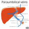
Data item |
Radiopaedia - Drawing Paraumbilical veins - English labels |
 |
rva |
Creative Commons Attribution-NonCommercial-ShareAlike |
1.141 |
2 |
|
Data item |
Sobotta 1909 fig.562 - Arteries and nerves of the palm of the hand - English labels |
 |
Student128 |
Public Domain |
1.141 |
2 |
|
Data item |
OLI - Drawing Cardiac conduction system and action potentials - English labels |
 |
rva |
Creative Commons Attribution-NonCommercial-ShareAlike |
1.140 |
1 |
|
Anatomical Structure |
Pars alveolaris mandibulae |
|
admin |
|
1.140 |
2 |
|
Data item |
Servier - Drawing Vertebral column lateral view - no labels |
 |
rva |
Creative Commons Attribution |
1.139 |
2 |
|
Anatomical Structure |
Ramus circumflexus (Arteria coronaria sinistra) |
|
admin |
|
1.138 |
2 |
|
Data item |
Anatomy Standard - Drawing Os scaphoideum - Latin labels |
 |
rva |
Creative Commons Attribution-NonCommercial |
1.137 |
4 |
|
Data item |
Anatomy Standard - Drawing Bony pelvis: differences between male and female (inferior view) - Latin labels |
 |
rva |
Creative Commons Attribution-NonCommercial |
1.136 |
2 |
|
Data item |
OpenStax AnatPhys fig.22.5 - Pseudostratified Epithelium - English labels |
 |
Jorn IJkhout |
Creative Commons Attribution |
1.136 |
4 |
|
Anatomical Structure |
Pars orbitalis ossis frontalis |
|
admin |
|
1.134 |
1 |
|
Data item |
U.Br.Columbia - Drawing Brain: lobes and landmarks - English labels |
 |
rva |
Creative Commons Attribution-NonCommercial-ShareAlike |
1.134 |
3 |
|
Data item |
Servier - Drawing Eye lateral view - no labels |
 |
rva |
Creative Commons Attribution |
1.134 |
4 |
|
Data item |
Slagter - Drawing Zenker's diverticulum - no labels |
 |
ProjectTulip |
Creative Commons Attribution-NonCommercial-ShareAlike |
1.134 |
1 |
|
Data item |
Anatomy Standard - Drawing Clavicula: inferior view - no labels |
 |
rva |
Creative Commons Attribution-NonCommercial |
1.133 |
1 |
|
Anatomical Structure |
Lamina lateralis processi pterygoideus ossis sphenoidalis |
|
admin |
|
1.133 |
4 |
|
Data item |
Anatomy Standard - Drawing Sacrum: superior aspect - Latin labels |
 |
rva |
Creative Commons Attribution-NonCommercial |
1.132 |
2 |
|
Learning Path |
Quiz Hart - Coronairvaten (basis) |
|
opgobee |
Creative Commons Attribution-NonCommercial-ShareAlike |
1.132 |
0 |
|
Data item |
Leiden, Maas - Presentation slides Pelvis plastination specimens PART 2 |
|
opgobee |
Creative Commons Attribution-NonCommercial-ShareAlike |
1.132 |
1 |
|
Data item |
Slagter - Drawing Larynx and vocal cords - no labels |
 |
rva |
Creative Commons Attribution-NonCommercial-ShareAlike |
1.132 |
4 |
|
Data item |
Slagter - Drawing Kidney macroscopic and microscopic anatomy - Dutch labels |
 |
rva |
Creative Commons Attribution-NonCommercial-ShareAlike |
1.132 |
1 |
|
Learning Path |
Quiz Thorax anatomie - Klinische toepassingen |
|
opgobee |
Creative Commons Attribution-NonCommercial-ShareAlike |
1.131 |
0 |
|
Data item |
Sobotta 1909 fig.770 - Orbital septum, anterior view - English labels |
 |
Student128 |
Public Domain |
1.130 |
2 |
|
Data item |
Prostate venous plexus, venous drainage penis and pudendal nerve – No labels |
 |
Siem Zethof |
Creative Commons Attribution-NonCommercial-ShareAlike |
1.129 |
2 |
|
Data item |
Sobotta 1909 fig.661 - Lamina quadrigemina and rhomboid fossa, lateral view - English labels |
 |
Student128 |
Public Domain |
1.129 |
1 |
|
Data item |
Lateral view of thorax with lung, skin, ribs and shoulder blade |
 |
Nadja Baltensweiler |
Creative Commons Attribution-NonCommercial-ShareAlike |
1.129 |
2 |
|
Data item |
Slagter - Drawing Brachial plexus and its relation to the muscles of arm - no labels |
 |
rva |
Creative Commons Attribution-NonCommercial-ShareAlike |
1.129 |
3 |
|
Data item |
Cartoon to remember the position of the structures in the hepatoduodenal ligament 2 - English labels |
 |
opgobee |
Creative Commons Attribution-NonCommercial-ShareAlike |
1.127 |
3 |
|
Data item |
RCSI - Drawing Nasopharyngeal arteries and Kiesselbach's plexus - English labels |
 |
rva |
Creative Commons Attribution-NonCommercial-ShareAlike |
1.125 |
5 |
|
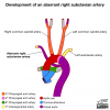
Data item |
Radiopaedia - Drawing Development of aberrant right subclavian artery - English labels |
 |
rva |
Creative Commons Attribution-NonCommercial-NoDerivs |
1.124 |
3 |
|
Anatomical Structure |
Arcus superciliaris |
|
admin |
|
1.124 |
1 |
|
Data item |
Anatomy Standard - Drawing Proximal ulna - Latin labels |
 |
rva |
Creative Commons Attribution-NonCommercial |
1.123 |
5 |
|
Anatomical Structure |
Ligamentum suspensorium ovarii |
|
admin |
|
1.123 |
0 |
|
Data item |
RCSI - Drawing Deep extensor muscles and tendons of forearm - English labels |
 |
rva |
Creative Commons Attribution-NonCommercial-ShareAlike |
1.123 |
3 |
|
Data item |
Sobotta 1906 fig.416 - Transverse section through the upper abdominal cavity - English labels |
 |
Student128 |
Public Domain |
1.123 |
2 |
|
Anatomical Structure |
Tuberositas deltoidea |
|
admin |
|
1.120 |
0 |
|
Data item |
Blausen - Bones of the skull - English labels |
 |
Student10 |
Creative Commons Attribution-ShareAlike |
1.118 |
1 |
|
Data item |
OpenStax AnatPhys fig.13.3 - Brain Vesicle Development - English labels |
 |
Jorn IJkhout |
Creative Commons Attribution |
1.118 |
1 |
|
Data item |
Sobotta 1909 fig.626 - Fissures and convulsions of the cerebral cortex, lateral view - coloured, no labels |
 |
rva |
Creative Commons Attribution-ShareAlike |
1.116 |
2 |
|
Anatomical Structure |
Vagina carotica |
|
admin |
|
1.115 |
2 |
|
Data item |
Froodrice - 3D model Circle of Willis |
|
rva |
Creative Commons Attribution-ShareAlike |
1.114 |
2 |
|
Data item |
Anatomy Standard - Drawing Sacrum: lateral aspect - Latin labels |
 |
rva |
Creative Commons Attribution-NonCommercial |
1.114 |
2 |
|
Learning Path |
Quiz Nier - Vascularisatie (basis) |
|
tjscherphof |
Creative Commons Attribution-NonCommercial-ShareAlike |
1.113 |
0 |
|
Anatomical Structure |
Ovarium |
|
admin |
|
1.112 |
1 |
|
Data item |
LadyofHats - Drawing Sutures of the cranium - Latin labels |
 |
rva |
Public Domain |
1.111 |
4 |
|
Data item |
Anatomy Standard - Drawing Medial view of 7th rib and vertebra - Latin labels |
 |
rva |
Creative Commons Attribution-NonCommercial |
1.111 |
1 |
|
Data item |
Radiopaedia - Drawing Rectus sheath above arcuate line - English labels |
 |
rva |
Creative Commons Attribution-NonCommercial-ShareAlike |
1.110 |
2 |
|
Anatomical Structure |
Facies lunata acetabuli |
|
admin |
|
1.109 |
2 |
|
Learning Path |
Liver Pancreas Gall bladder - Entrance exam |
|
leo.koehler |
Creative Commons Attribution-NonCommercial-ShareAlike |
1.107 |
1 |
|
Data item |
Leiden - Drawing The cardiac conduction system - no labels |
 |
rva |
Creative Commons Attribution-NonCommercial-ShareAlike |
1.106 |
2 |
|
Data item |
Sobotta 1909 fig.154 - bones of the foot, dorsal surface - English Labels |
 |
Student128 |
Public Domain |
1.104 |
2 |
|
Data item |
Anatomy Standard - Drawing Thorcacic vertebra (Th5): anterior view - Latin labels |
 |
rva |
Creative Commons Attribution-NonCommercial |
1.104 |
3 |
|
Anatomical Structure |
Crista nasalis maxillae |
|
admin |
|
1.103 |
0 |
|
Learning Path |
Quiz Nier - Kenmerken Inwendig (gevorderd) |
|
tjscherphof |
Creative Commons Attribution-NonCommercial-ShareAlike |
1.102 |
0 |
|
Anatomical Structure |
Truncus pulmonalis |
|
admin |
|
1.101 |
2 |
|
Data item |
Borgquist - Drawing Coronary veins from a posterior view and an anterior view - English labels |
 |
opgobee |
Creative Commons Attribution |
1.101 |
5 |
|
Data item |
BlueLink - 3D model Right Coxal bone |
|
rva |
Creative Commons Attribution-NonCommercial-NoDerivs |
1.098 |
3 |
|
Data item |
Servier - Drawing Bronchial tree - no labels |
 |
rva |
Creative Commons Attribution |
1.098 |
1 |
|
Anatomical Structure |
Os palatinum |
|
admin |
|
1.097 |
1 |
|
Anatomical Structure |
Cavea thoracis |
|
admin |
|
1.097 |
0 |
|
Data item |
Inferior view of the female pelvic diaphragm - no labels |
 |
admin |
Creative Commons Attribution-NonCommercial-ShareAlike |
1.096 |
1 |
|
Data item |
Lynch - Drawing Lateral view of the brain in the skull - no labels |
 |
rva |
Creative Commons Attribution |
1.095 |
2 |
|
Data item |
Leiden - Student - Drawing Coronary vessels - Numbered labels and Latin answers |
 |
Flynn Post |
Creative Commons Attribution-ShareAlike |
1.094 |
2 |
|
Anatomical Structure |
Facies externa ossis frontalis |
|
admin |
|
1.094 |
3 |
|
Data item |
OLI - Drawing Structure of skeletal muscle - English labels |
 |
rva |
Creative Commons Attribution-NonCommercial-ShareAlike |
1.093 |
1 |
|
Data item |
Inferior view of the male pelvic diaphragm - English labels |
 |
opgobee |
Creative Commons Attribution-NonCommercial-ShareAlike |
1.092 |
1 |
|
Learning Path |
Demonstratie Anatomie Mediastinum en Pericard |
|
FrisoJansen |
This item is (on) an external site. The license as stated on that site holds. |
1.091 |
0 |
|
Data item |
Sobotta 1909 fig.588 - Great venous and arterial trunks of the thorax and deep vessels of the neck - English labels |
 |
Student128 |
Public Domain |
1.091 |
2 |
|
Learning Path |
Quiz Anatomie van Pijn B - Routes en Referred Pain 2 |
|
opgobee |
Creative Commons Attribution-NonCommercial-ShareAlike |
1.091 |
1 |
|
Data item |
BlueLink - 3D model Eleventh Thoracic Vertebra (T11) |
|
rva |
Creative Commons Attribution-NonCommercial-NoDerivs |
1.091 |
1 |
|
Data item |
Slagter - Drawing Inferior hypogastric plexus and pudendal nerve, male - No labels |
 |
Siem Zethof |
Creative Commons Attribution-NonCommercial-ShareAlike |
1.089 |
1 |
|
Learning Path |
Quiz Anatomie van Pijn C - Klinische toepassingen 2 |
|
opgobee |
Creative Commons Attribution-NonCommercial-ShareAlike |
1.088 |
1 |
|
Data item |
Leiden - Drawing pancreas and neighbouring organs - no labels |
 |
opgobee |
Creative Commons Attribution-NonCommercial-ShareAlike |
1.088 |
3 |
|
Anatomical Structure |
Corpus gastricum |
|
admin |
|
1.088 |
2 |
|
Data item |
Anatomy Standard - Drawing Distal fibula: anterior and posterior view - Latin labels |
 |
rva |
Creative Commons Attribution-NonCommercial |
1.088 |
2 |
|
Data item |
Sobotta 1909 fig.763 - The tarsal plates with palpebral ligaments and lachrymal sac - English labels |
 |
Student128 |
Public Domain |
1.088 |
3 |
|
Learning Path |
Quiz Hart - Kleppen (gevorderd) |
|
opgobee |
Creative Commons Attribution-NonCommercial-ShareAlike |
1.088 |
0 |
|
Data item |
KnowledgeWorks - Drawing Kidney anatomy and detail - English labels |
 |
rva |
Creative Commons Attribution |
1.087 |
1 |
|
Data item |
U.Br.Columbia - Drawing The cubital fossa - English labels |
 |
rva |
Creative Commons Attribution-NonCommercial-ShareAlike |
1.086 |
1 |
|
Data item |
Radiopaedia - Drawing Anatomy of the knee from posterior: bones - English labels |
 |
rva |
Creative Commons Attribution-NonCommercial-ShareAlike |
1.085 |
2 |