
nid: 62437
Additional formats:
None available
Description:
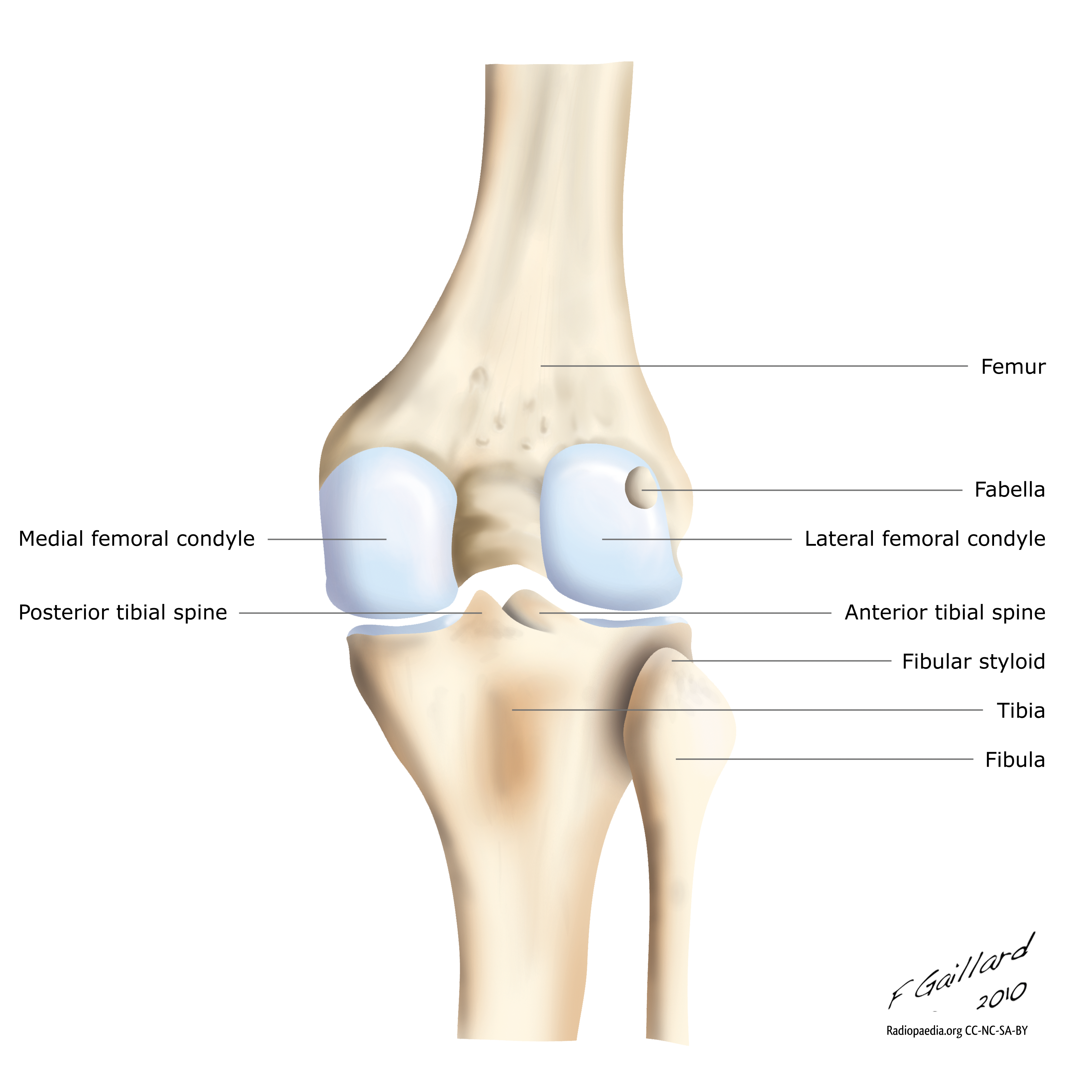
Anatomy of the knee from posterior: bones. English labels.
Case courtesy of Assoc Prof Frank Gaillard, Radiopaedia.org. From the case rID: 9330
Case courtesy of Assoc Prof Frank Gaillard, Radiopaedia.org. From the case rID: 9330
Anatomical structures in item:
Uploaded by: rva
Netherlands, Leiden – Leiden University Medical Center, Leiden University
Genu
Femur
Tibia
Fibula
Condylus lateralis femoris
Condylus medialis femoris
Creator(s)/credit: Dr Frank Gaillard MB.BS, MMed
Requirements for usage
You are free to use this item if you follow the requirements of the license:  View license
View license If you use this item you should credit it as follows:
- For usage in print - copy and paste the line below:
- For digital usage (e.g. in PowerPoint, Impress, Word, Writer) - copy and paste the line below (optionally add the license icon):
"Radiopaedia - Drawing Anatomy of the knee from posterior: bones - English labels" at AnatomyTOOL.org by Frank Gaillard, license: Creative Commons Attribution-NonCommercial-ShareAlike


Comments