|
Data item |
Leiden MOOC 3.7 embryonic development of the gut, an animation |
|
rjjvisser |
Creative Commons Attribution-NonCommercial-ShareAlike |
1.417 |
3 |
|
Data item |
Servier - Drawing Neuron - no labels |
 |
rva |
Creative Commons Attribution |
1.416 |
4 |
|
Data item |
U.Br.Columbia - Drawing Superficial anatomy of the thigh - English labels |
 |
rva |
Creative Commons Attribution-NonCommercial-ShareAlike |
1.416 |
2 |
|
Data item |
Slagter - Drawing Hepatic segments - Dutch labels |
 |
rva |
Creative Commons Attribution-NonCommercial-ShareAlike |
1.416 |
3 |
|
Data item |
U.Br.Columbia - Drawing The nerves in the cranial fossae - English labels |
 |
rva |
Creative Commons Attribution-NonCommercial-ShareAlike |
1.414 |
2 |
|
Data item |
Anatomy Standard - Drawing Scapula: lateral view - no labels |
 |
rva |
Creative Commons Attribution-NonCommercial |
1.412 |
2 |
|
Data item |
OpenStax AnatPhys fig.7.27 - Thoracic Vertebra and Rib - English labels |
 |
Jorn IJkhout |
Creative Commons Attribution |
1.410 |
3 |
|
Data item |
NYSORA - Drawing Cross-section adductors thigh - English labels |
 |
rva |
Creative Commons Attribution-NonCommercial-NoDerivs |
1.409 |
1 |
|
Anatomical Structure |
Tuberositas pterygoidea |
|
admin |
|
1.408 |
3 |
|
Data item |
Dundee - Diagram Major peripheral nervous system nerves - English labels |
 |
rva |
Creative Commons Attribution-NonCommercial-NoDerivs |
1.407 |
1 |
|
Anatomical Structure |
Ventriculus |
|
admin |
|
1.406 |
4 |
|
Data item |
OLI - Drawing Circle of Willis - English labels |
 |
rva |
Creative Commons Attribution-NonCommercial-ShareAlike |
1.406 |
4 |
|
Data item |
Leiden - Drawing Liver segments and vascularisation - Dutch/Latin labels |
 |
opgobee |
Creative Commons Attribution-NonCommercial-ShareAlike |
1.406 |
5 |
|
Data item |
Lynch - Drawing Anterior and lateral view of skeleton - no labels |
 |
rva |
Creative Commons Attribution |
1.403 |
1 |
|
Anatomical Structure |
Juga alveolaria mandibulae |
|
admin |
|
1.402 |
4 |
|
Data item |
Slagter - Drawing Larynx anatomy: anterior lateral view - no labels |
 |
rva |
Creative Commons Attribution-NonCommercial-ShareAlike |
1.401 |
2 |
|
Data item |
Cenveo - Drawing Lateral view of the brain - English labels |
 |
rva |
Creative Commons Attribution |
1.401 |
4 |
|
Data item |
Dundee - Drawing Abduction of the hip: origin and insertio of tensor fasciae latae - English labels |
 |
rva |
Creative Commons Attribution-NonCommercial-NoDerivs |
1.400 |
2 |
|
Data item |
U.Br.Columbia - Drawing Origins and insertions of the extrinsic tongue muscles - English labels |
 |
rva |
Creative Commons Attribution-NonCommercial-ShareAlike |
1.400 |
2 |
|
Data item |
U.Br.Columbia - Drawing Place of the heart in thorax and auscultation of the valves - English labels |
 |
rva |
Creative Commons Attribution-NonCommercial-ShareAlike |
1.399 |
1 |
|
Data item |
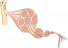
Slagter - Drawing Stages of fertilisation of oocyte - English labels |
 |
rva |
Creative Commons Attribution-NonCommercial-ShareAlike |
1.398 |
3 |
|
Data item |
Leiden - Presentation slide Photo internal view female inguinal region |
|
opgobee |
Creative Commons Attribution-NonCommercial-ShareAlike |
1.398 |
1 |
|
Anatomical Structure |
Basis cranii |
|
admin |
|
1.397 |
1 |
|
Data item |
Anatomy Standard - Drawing Right ulna: anterior view - Latin labels |
 |
rva |
Creative Commons Attribution-NonCommercial |
1.396 |
5 |
|
Data item |
Radiopaedia - Drawing Branches of the abdominal aorta - English labels |
 |
rva |
Creative Commons Attribution-NonCommercial-ShareAlike |
1.391 |
2 |
|
Anatomical Structure |
Recessus piriformis |
|
admin |
|
1.390 |
1 |
|
Anatomical Structure |
Sulcus obturatorius |
|
admin |
|
1.388 |
3 |
|
Data item |
RCSI - Drawing Arteries and veins of kidney and adrenal gland - English labels |
 |
opgobee |
Creative Commons Attribution-NonCommercial-ShareAlike |
1.387 |
2 |
|
Anatomical Structure |
Facies dorsalis ossis sacri |
|
admin |
|
1.385 |
4 |
|
Data item |
Slagter - Drawing Large intestine with vascularisation and mesocolons - No labels |
 |
lumcanatomy |
Creative Commons Attribution-NonCommercial-ShareAlike |
1.385 |
3 |
|
Data item |
KnowledgeWorks - Drawing Location of salivary glands and ducts - English labels |
 |
rva |
Creative Commons Attribution |
1.384 |
4 |
|
Anatomical Structure |
Ramus interventricularis anterior (Arteria coronaria sinistra) |
|
admin |
|
1.384 |
1 |
|
Data item |
OpenStax AnatPhys fig.7.18 - Paranasal Sinuses - English labels |
 |
Jorn IJkhout |
Creative Commons Attribution |
1.382 |
4 |
|
Data item |
U.Br.Columbia - Drawing Cross section through middle of leg - English labels |
 |
rva |
Creative Commons Attribution-NonCommercial-ShareAlike |
1.382 |
1 |
|
Learning Path |
Quiz Female Internal Genital Organs (advanced) |
|
EmmaL |
Creative Commons Attribution-NonCommercial-ShareAlike |
1.380 |
0 |
|
Data item |
Sobotta 1909 fig.563 - Arteries and nerves of the palm of the hand - English labels |
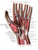 |
Student128 |
Public Domain |
1.380 |
2 |
|
Anatomical Structure |
Angulus sterni |
|
admin |
|
1.379 |
0 |
|
Data item |
Radiopaedia - Drawing Talus from multiple angles - English labels |
 |
rva |
Creative Commons Attribution-NonCommercial-NoDerivs |
1.379 |
3 |
|
Data item |
Sobotta 1909 fig.646 - Frontal section of the brain - English labels |
 |
Student128 |
Public Domain |
1.379 |
3 |
|
Data item |
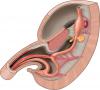
Leiden - Drawing Innervation of female genitalia - English labels |
 |
rva |
Creative Commons Attribution-NonCommercial-ShareAlike |
1.378 |
3 |
|
Anatomical Structure |
Facies anterior partis petrosae ossis temporalis |
|
admin |
|
1.377 |
0 |
|
Data item |
3D Anatomy Lyon: Muscles for adduction of the arm - video of 3D model |
|
rva |
Creative Commons Attribution-NonCommercial-NoDerivs |
1.376 |
3 |
|
Data item |
Leiden - Median laparotomy incision - no labels |
 |
opgobee |
Creative Commons Attribution-NonCommercial-ShareAlike |
1.375 |
1 |
|
Data item |
Leiden - Photo Mesentery of the small intestine - no labels |
 |
opgobee |
Creative Commons Attribution-NonCommercial-ShareAlike |
1.375 |
4 |
|
Data item |
Slagter - Drawing Stomach, pancreas, duodenum and liver - no labels |
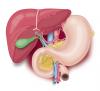 |
rva |
Creative Commons Attribution-NonCommercial-ShareAlike |
1.374 |
2 |
|
Anatomical Structure |
Cingulum pectorale |
|
admin |
|
1.374 |
2 |
|
Data item |
Sobotta 1909 fig.306 - anterior muscles of the leg - no labels |
 |
lumcanatomy |
Creative Commons Attribution-ShareAlike |
1.373 |
1 |
|
Data item |
OpenStax AnatPhys fig.8.19 - Bones of the Foot - English labels |
 |
Jorn IJkhout |
Creative Commons Attribution |
1.373 |
7 |
|
Data item |
OpenStax AnatPhys fig.20.29 - Thoracic Abdominal Arteries Chart - English labels |
 |
Jorn IJkhout |
Creative Commons Attribution |
1.371 |
1 |
|
Anatomical Structure |
Tuber omentale pancreatis |
|
admin |
|
1.370 |
3 |
|
Data item |
Slagter - Drawing Anatomy of kidney, nephron and glomerulus - no labels |
 |
rva |
Creative Commons Attribution-NonCommercial-ShareAlike |
1.368 |
4 |
|
Data item |
Leiden MOOC 5.7 - Video Anatomy on the table: demonstration of the deep body wall |
|
rjjvisser |
Creative Commons Attribution-NonCommercial-ShareAlike |
1.366 |
1 |
|
Data item |
Anatomy Standard - Drawing Costovertebral joints: first and eight rib (ex situ) - Latin labels |
 |
rva |
Creative Commons Attribution-NonCommercial |
1.365 |
4 |
|
Data item |
Cenveo - Drawing Anatomy of the Cochlea - English labels |
 |
rva |
Creative Commons Attribution |
1.363 |
3 |
|
Data item |
Palmer - Drawing Cross-section of testis - English labels |
 |
rva |
Creative Commons Attribution |
1.359 |
6 |
|
Data item |
StatPearls - Drawing Branches of the posterior cerebral artery - English labels |
 |
rva |
Creative Commons Attribution-NonCommercial-NoDerivs |
1.359 |
3 |
|
Anatomical Structure |
Incisura trochlearis |
|
admin |
|
1.359 |
3 |
|
Data item |
Nahabedian - Drawing Internal view of left ventricle - English labels |
 |
rva |
Creative Commons Attribution-NonCommercial-NoDerivs |
1.358 |
1 |
|
Data item |
Sobotta 1909 fig.726 - Sympathetic trunk and vagus nerve, thoracic (and abdominal) portion - English labels |
 |
Student128 |
Public Domain |
1.357 |
2 |
|
Data item |
Dundee - Drawing Course of the Facial Nerve within the Inner Ear - English labels |
 |
rva |
Creative Commons Attribution-NonCommercial-NoDerivs |
1.355 |
4 |
|
Data item |
Radiopaedia - Drawing Contents of the cavernous sinus - English labels |
 |
rva |
Creative Commons Attribution-NonCommercial-ShareAlike |
1.354 |
4 |
|
Anatomical Structure |
Arcus anterior atlantis |
|
admin |
|
1.354 |
0 |
|
Data item |
Slagter - Drawing Inferior view vulva and anus female - no labels |
 |
opgobee |
Creative Commons Attribution-NonCommercial-ShareAlike |
1.352 |
4 |
|
Data item |
Anterior view of the abdomen with the dotted line indicating the line of Toldt of the colon descendens – no labels |
 |
Siem Zethof |
Creative Commons Attribution-NonCommercial-ShareAlike |
1.350 |
7 |
|
Data item |
MedicalGraphics - Drawing Open mouth with tongue and teeth - no labels |
 |
rva |
Creative Commons Attribution-NoDerivatives |
1.349 |
2 |
|
Data item |
Groningen - 3D model Cardiac Conduction System and Coronaries |
|
rva |
Creative Commons Attribution-NonCommercial-ShareAlike |
1.348 |
3 |
|
Anatomical Structure |
Sulcus arteriae occipitalis |
|
admin |
|
1.348 |
0 |
|
Anatomical Structure |
Os frontale |
|
admin |
|
1.344 |
2 |
|
Data item |
Anatomy Standard - Drawing Anatomy of the distal femur - no labels |
 |
rva |
Creative Commons Attribution-NonCommercial |
1.344 |
2 |
|
Data item |
Radiopaedia - Drawing Midbrain at level of trochlear nerve - English labels |
 |
rva |
Creative Commons Attribution-NonCommercial-ShareAlike |
1.344 |
2 |
|
Data item |
Blausen 0020 - Landmarks of surface anatomy (Anterior view) - English labels |
 |
Student10 |
Creative Commons Attribution |
1.343 |
3 |
|
Anatomical Structure |
Plica salpingopalatina |
|
admin |
|
1.342 |
5 |
|
Anatomical Structure |
Aorta |
|
admin |
|
1.341 |
1 |
|
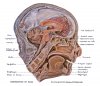
Data item |
Radiopaedia - Drawing Base of skull - English labels |
 |
rva |
Creative Commons Attribution-NonCommercial-ShareAlike |
1.340 |
2 |
|
Data item |
Slagter - Drawing Anatomy of the fundus of the eye - Dutch labels |
 |
rva |
Creative Commons Attribution-NonCommercial-ShareAlike |
1.338 |
4 |
|
Data item |
Anatomy Standard - Drawing Superior aspect of cervical vertebra (C4) - no labels |
 |
rva |
Creative Commons Attribution-NonCommercial |
1.337 |
5 |
|
Data item |
NYSORA - Drawing Nerves of pelvis - English labels |
 |
rva |
Creative Commons Attribution-NonCommercial-NoDerivs |
1.336 |
2 |
|
Data item |
U.Br.Columbia - Drawing Parasagittal section of head - English labels |
 |
rva |
Creative Commons Attribution-NonCommercial-ShareAlike |
1.335 |
3 |
|
Data item |
Groningen - 3D model Anatomy of the mandible and adjacent structures in sagittal cut |
|
rva |
Creative Commons Attribution-NonCommercial-ShareAlike |
1.334 |
3 |
|
Data item |
Greensboro - 3D model Sternum with sternal foramen |
|
rva |
Creative Commons Attribution |
1.334 |
2 |
|
Data item |
Cenveo - Drawing Superior view of the brain - English labels |
 |
rva |
Creative Commons Attribution |
1.333 |
4 |
|
Data item |
Sobotta 1909 fig.716 - Lumbal plexus and blood vessels of the posterior abdominal wall - English labels |
 |
Student128 |
Public Domain |
1.331 |
1 |
|
Data item |
Syndey - 3D model Oral Cavity |
|
rva |
Creative Commons Attribution-ShareAlike |
1.329 |
2 |
|
Anatomical Structure |
Squama frontalis |
|
admin |
|
1.329 |
2 |
|
Data item |
Leiden, Maas - Presentation slides Pelvis plastination specimens PART 1 |
|
opgobee |
Creative Commons Attribution-NonCommercial-ShareAlike |
1.328 |
2 |
|
Data item |
Anatomy Standard - Drawing Axis: lateral aspect - Latin labels |
 |
rva |
Creative Commons Attribution-NonCommercial |
1.328 |
3 |
|
Anatomical Structure |
Rima pudendi |
|
admin |
|
1.327 |
2 |
|
Data item |
Servier - Drawing Structure of the skeletal muscle - no labels |
 |
rva |
Creative Commons Attribution |
1.327 |
6 |
|
Anatomical Structure |
Plica spiralis |
|
admin |
|
1.325 |
0 |
|
Learning Path |
Quiz Hart - Coronairvaten op coronairangiografiën |
|
opgobee |
Creative Commons Attribution-NonCommercial-ShareAlike |
1.325 |
1 |
|
Data item |
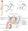
OpenStax AnatPhys fig.27.2 - Male Reproductive System - English labels |
 |
Jorn IJkhout |
Creative Commons Attribution |
1.325 |
2 |
|
Data item |
Leiden - Drawing Stomach, duodenum, pancreas - no labels |
 |
opgobee |
Creative Commons Attribution-NonCommercial-ShareAlike |
1.323 |
3 |
|
Data item |
Slagter - Drawing Spermatogenesis in seminiferous tubules of testis - no labels |
 |
rva |
Creative Commons Attribution-NonCommercial-ShareAlike |
1.323 |
3 |
|
Data item |
OpenStax AnatPhys fig.7.6 - Cranial Fossae - English labels |
 |
Jorn IJkhout |
Creative Commons Attribution |
1.321 |
2 |
|
Data item |
Radiopaedia - Drawing Development of the aortic arch and branches at 7 weeks - English labels |
 |
rva |
Creative Commons Attribution-NonCommercial-NoDerivs |
1.320 |
1 |
|
Data item |
OpenStax AnatPhys fig.16.15 - Major Regions of the Cerebellum - English labels |
 |
Jorn IJkhout |
Creative Commons Attribution |
1.320 |
3 |
|
Data item |
Drawing digestive system in embryo |
 |
lumcanatomy |
Creative Commons Attribution-NonCommercial-ShareAlike |
1.317 |
2 |
|
Data item |
Groningen - 3D model Lymphatic system: anatomy of a lymph node |
|
rva |
Creative Commons Attribution-NonCommercial-ShareAlike |
1.315 |
4 |
|
Anatomical Structure |
Corpus ossis sphenoidalis |
|
admin |
|
1.315 |
4 |
|
Data item |
Sobotta 1914 fig.536-537 - Coronary arteries and veins - English labels |
 |
Student128 |
Public Domain |
1.314 |
3 |