
nid: 58317
Additional formats:
None available
Description:
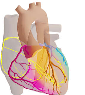
Cardiac Conduction System and Coronaries. This 3D model shows the cardiac conduction system and the coronaries, in the canonical configuration of the coronaries. The coronaries have been given colours. Corresponding faint transparent colour shadings indicate the supplied cardiac areas by each coronary. It can be observed that the dorsally lying AV node is supplied by a branch from the right coronary, whilst the bundle branches that lie centrally in the septum are in the left coronary's supply area.
fThe conduction system of the heart consists of cardiac muscle cells and conducting fibers (not nervous tissue) that are specialized for initiating impulses and conducting them rapidly through the heart. They initiate the normal cardiac cycle and coordinate the contractions of cardiac chambers. The conducting system provides the heart its automatic rhythmic beat. For the heart to pump efficiently and the systemic and pumonary circulations to operate in synchrony, the events in the cardiac cycle must be coordinated.
fThe conduction system of the heart consists of cardiac muscle cells and conducting fibers (not nervous tissue) that are specialized for initiating impulses and conducting them rapidly through the heart. They initiate the normal cardiac cycle and coordinate the contractions of cardiac chambers. The conducting system provides the heart its automatic rhythmic beat. For the heart to pump efficiently and the systemic and pumonary circulations to operate in synchrony, the events in the cardiac cycle must be coordinated.
Anatomical structures in item:
Uploaded by: rva
Netherlands, Leiden – Leiden University Medical Center, Leiden University
Nodus sinuatrialis
Nodus atrioventricularis
Fasciculus atrioventricularis
Rami subendocardiales fasciculi atrioventricularis
Cor
Creator(s)/credit: Anna Sieben
Requirements for usage
You are free to use this item if you follow the requirements of the license:  View license
View license If you use this item you should credit it as follows:
- For usage in print - copy and paste the line below:
- For digital usage (e.g. in PowerPoint, Impress, Word, Writer) - copy and paste the line below (optionally add the license icon):
"Groningen - 3D model Cardiac Conduction System and Coronaries" at AnatomyTOOL.org by Anna Sieben, license: Creative Commons Attribution-NonCommercial-ShareAlike. From E-learning UMCG https://sketchfab.com/eLearningUMCG
"Groningen - 3D model Cardiac Conduction System and Coronaries" by Anna Sieben, license: CC BY-NC-SA. From E-learning UMCG https://sketchfab.com/eLearningUMCG


Comments