
nid: 60125
Additional formats:
None available
Description:
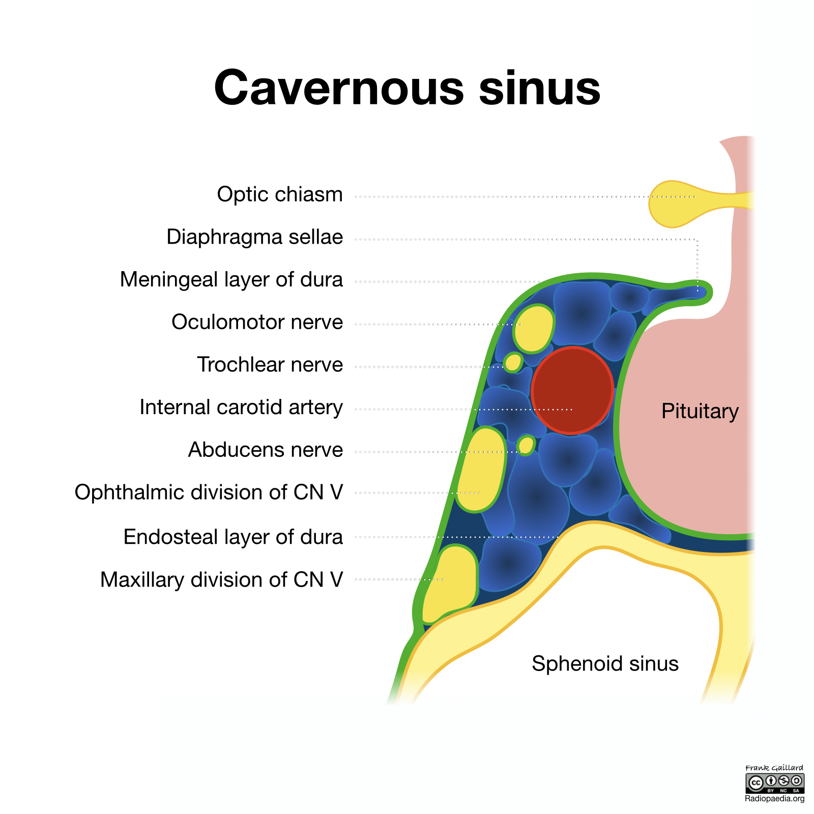
Contents of the cavernous sinus. This coronal section of the cavernous sinus shows the structures that pass through it. English labels.
Case courtesy of Assoc Prof Frank Gaillard, Radiopaedia.org. From the case rID: 54907
Case courtesy of Assoc Prof Frank Gaillard, Radiopaedia.org. From the case rID: 54907
Anatomical structures in item:
Uploaded by: rva
Netherlands, Leiden – Leiden University Medical Center, Leiden University
Sinus cavernosus
Glandula pituitaria
Sinus sphenoidalis
Chiasma opticum
Diaphragma sellae
Nervus oculomotorius [III]
Nervus trochlearis [IV]
Arteria carotis interna
Nervus abducens [VI]
Nervus ophthalmicus [Va]
Nervus maxillaris [Vb]
Creator(s)/credit: Frank Gaillard MB.BS, MMed
Requirements for usage
You are free to use this item if you follow the requirements of the license:  View license
View license If you use this item you should credit it as follows:
- For usage in print - copy and paste the line below:
- For digital usage (e.g. in PowerPoint, Impress, Word, Writer) - copy and paste the line below (optionally add the license icon):
"Radiopaedia - Drawing Contents of the cavernous sinus - English labels" at AnatomyTOOL.org by Frank Gaillard, license: Creative Commons Attribution-NonCommercial-ShareAlike


Comments