
nid: 60413
Additional formats:
None available
Description:
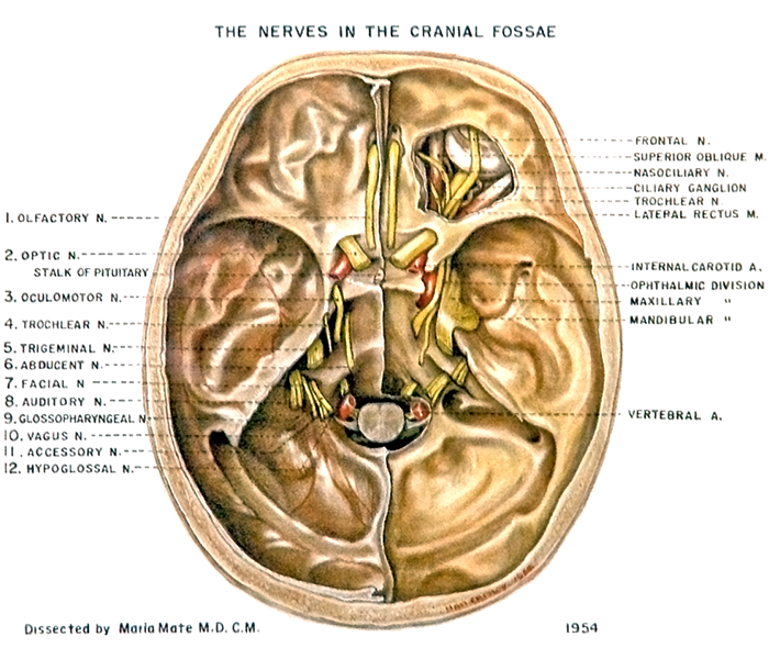
The nerves in the cranial fossae. The nerves in the basel of the skull can be seen in this image. English labels.
Retrieved from website Clinical Anatomy of the University of British Columbia.
Retrieved from website Clinical Anatomy of the University of British Columbia.
Anatomical structures in item:
Uploaded by: rva
Netherlands, Leiden – Leiden University Medical Center, Leiden University
Fila olfactoria
Nervus opticus
Nervus cranialis
Nervus oculomotorius [III]
Nervus trochlearis [IV]
Nervus trigeminus [V]
Nervus abducens [VI]
Nervus facialis [VII]
Nervus cochlearis
Nervus glossopharyngeus [IX]
Nervus vagus
Nervus accessorius [XI]
Nervus hypoglossus [XII]
Arteria vertebralis
Basis cranii
Arteria carotis interna
Nervus ophthalmicus [Va]
Nervus maxillaris [Vb]
Nervus mandibularis [Vc]
Ganglion ciliare
Nervus nasociliaris
Nervus frontalis
Creator(s)/credit: A.G.L. (Nan) Cheney, medical illustrator, UBC; Maria Mate MD, CM, UBC
Requirements for usage
You are free to use this item if you follow the requirements of the license:  View license
View license If you use this item you should credit it as follows:
- For usage in print - copy and paste the line below:
- For digital usage (e.g. in PowerPoint, Impress, Word, Writer) - copy and paste the line below (optionally add the license icon):
"U.Br.Columbia - Drawing The nerves in the cranial fossae - English labels" at AnatomyTOOL.org by A.G.L. (Nan) Cheney, UBC and Maria Mate, UBC, license: Creative Commons Attribution-NonCommercial-ShareAlike. Source: website Clinical Anatomy, http://www.clinicalanatomy.ca
"U.Br.Columbia - Drawing The nerves in the cranial fossae - English labels" by A.G.L. (Nan) Cheney, UBC and Maria Mate, UBC, license: CC BY-NC-SA. Source: website Clinical Anatomy, http://www.clinicalanatomy.ca


Comments