
nid: 59564
Additional formats:
None available
Description:
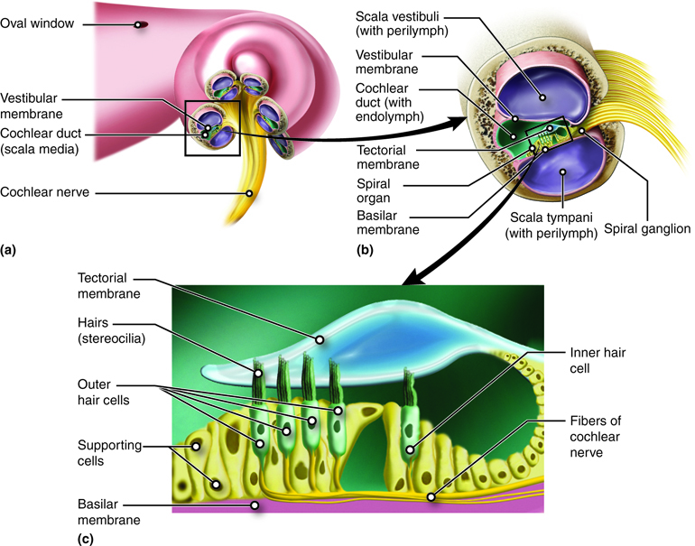
Anatomy of the Cochlea. The cochlea is a spiral structure (a) divided into three chambers (b). The middle chamber, the cochlear duct, contains the spiral organ that has hair cells (c) for sensing the vibrations we perceive as sound. English labels.
Anatomical structures in item:
Uploaded by: rva
Netherlands, Leiden – Leiden University Medical Center, Leiden University
Cochlea
Scala vestibuli
Scala tympani
Ganglion cochleare
Organum spirale
Membrana tectoria
Perilympha
Fossula fenestrae vestibuli
Creator(s)/credit: Cenveo
Requirements for usage
You are free to use this item if you follow the requirements of the license:  View license
View license If you use this item you should credit it as follows:
- For usage in print - copy and paste the line below:
- For digital usage (e.g. in PowerPoint, Impress, Word, Writer) - copy and paste the line below (optionally add the license icon):
"Cenveo - Drawing Anatomy of the Cochlea - English labels" at AnatomyTOOL.org by Cenveo, license: Creative Commons Attribution
"Cenveo - Drawing Anatomy of the Cochlea - English labels" by Cenveo, license: CC BY


Comments