
nid: 63090
Additional formats:
None available
Description:
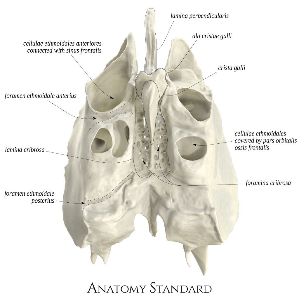
Ethnoid bone: superior view. The ethmoid bone is localized between the orbits and is a significant part of the nasal cavity. It is an unpaired bone, made almost entirely by thin bony lamellae. Note that multiple ethmoid cells open on top of the etmoidal labyrinth. The most ventral ethmoid cells communicate with the frontal sinus, and the dorsal ones are covered by the pars orbitalis ossis frontalis. The foramen ethmoidale anterius localized on the medial wall of the orbit connects the orbit with the fossa cranii anterior via a channel bordered by the ethmoid bone (channel's floor) and the frontal bone (the channel's roof). This channel is known as canalis ethmoidalis anterior, is about 6 mm long, and contains clinically important a. ethmoidalis anterior. Latin labels.
Image and description retrieved from Anatomy Standard. Via this link more images can be found, including oblique views.
Image and description retrieved from Anatomy Standard. Via this link more images can be found, including oblique views.
Anatomical structures in item:
Uploaded by: rva
Netherlands, Leiden – Leiden University Medical Center, Leiden University
Os ethmoidale
Lamina perpendicularis ossis ethmoidalis
Crista galli
Labyrinthus ethmoidalis
Cellulae ethmoidales
Cellulae ethmoidales anteriores
Cellulae ethmoidales mediae
Cellulae ethmoidales posteriores
Sinus frontalis
Foramen ethmoidale anterius
Lamina cribrosa
Foramen ethmoidale posterius
Foramina cribrosa
Ala cristae galli
Creator(s)/credit: Jānis Šavlovskis MD, PhD, Assistant Professor; Kristaps Raits, 3D generalist
Requirements for usage
You are free to use this item if you follow the requirements of the license:  View license
View license If you use this item you should credit it as follows:
- For usage in print - copy and paste the line below:
- For digital usage (e.g. in PowerPoint, Impress, Word, Writer) - copy and paste the line below (optionally add the license icon):
"Anatomy Standard - Drawing Ethmoid bone: superior view - Latin labels" at AnatomyTOOL.org by Jānis Šavlovskis and Kristaps Raits, license: Creative Commons Attribution-NonCommercial


Comments