
nid: 59625
Additional formats:
None available
Description:
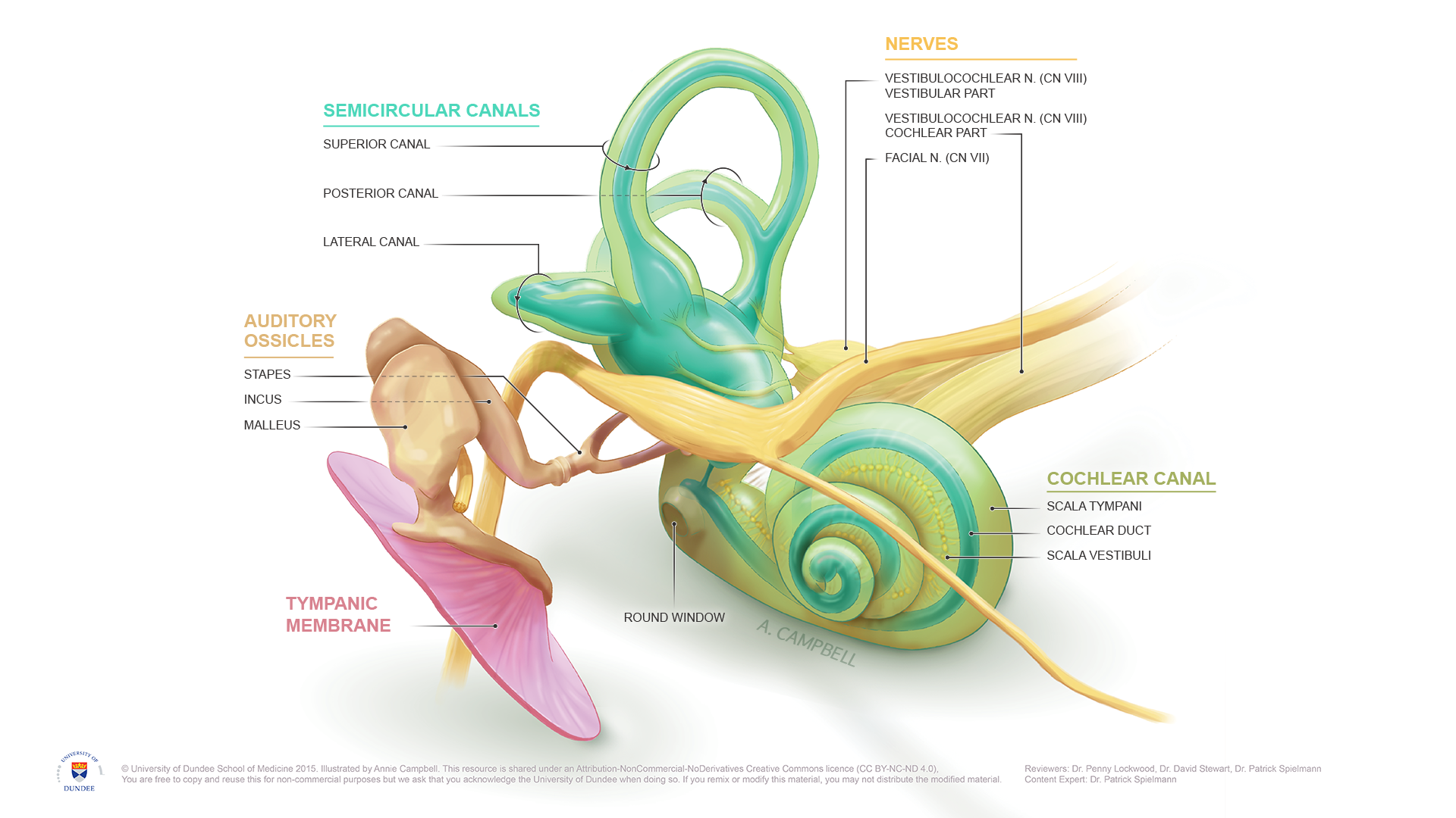
Anatomy of the Inner Ear. The inner ear is the interior part of the ear, it is important for the detection of sound an balance. English labels. NOTE: THIS IMAGE IS UNDER A NON-DERIVATIVE LICENSE. THIS MEANS THAT IF YOU REMIX OR REVISE THIS MATERIAL YOU MAY NOT DISTRIBUTE THE MODIFIED MATERIAL.
Anatomical structures in item:
Uploaded by: rva
Netherlands, Leiden – Leiden University Medical Center, Leiden University
Auris interna
Canales semicirculares
Canalis semicircularis anterior
Canalis semicircularis posterior
Canalis semicircularis lateralis
Ossicula auditoria
Stapes
Incus
Malleus
Membrana tympanica
Fenestra cochleae
Canaliculus cochleae
Scala tympani
Ductus cochlearis
Scala vestibuli
Nervus vestibulocochlearis [VIII]
Nervus vestibularis
Nervus cochlearis
Nervus facialis [VII]
Creator(s)/credit: Annie Campbell MSc, medical illustrator
Requirements for usage
You are free to use this item if you follow the requirements of the license:  View license
View license If you use this item you should credit it as follows:
- For usage in print - copy and paste the line below:
- For digital usage (e.g. in PowerPoint, Impress, Word, Writer) - copy and paste the line below (optionally add the license icon):
"Dundee - Drawing Anatomy of the Inner Ear - English labels" at AnatomyTOOL.org by Annie Campbell, © University of Dundee School of Medicine, license: Creative Commons Attribution-NonCommercial-NoDerivs. Reviewed by: Dr. Penny Lockwood, Dr. David Stewart, Dr. Patrick Spielmann
"Dundee - Drawing Anatomy of the Inner Ear - English labels" by Annie Campbell, © University of Dundee School of Medicine, license: CC BY-NC-ND. Reviewed by: Dr. Penny Lockwood, Dr. David Stewart, Dr. Patrick Spielmann


Comments