
nid: 63381
Additional formats:
None available
Description:
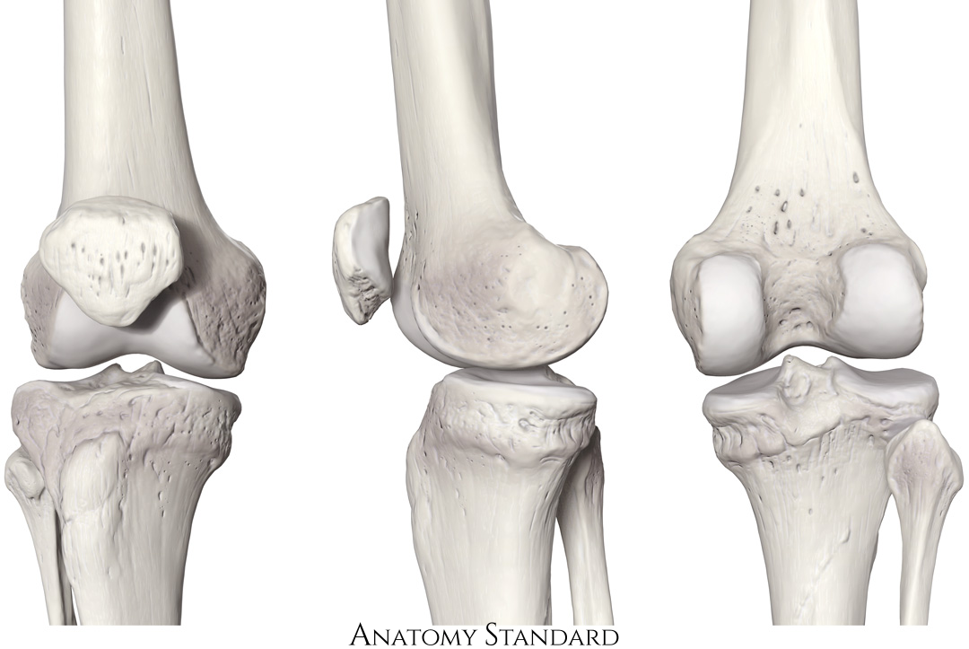
Knee joint and patella: anterior, medial and posterior view. This image shows the knee joint and patella from ventral, medial and dorsal. Version without labels.
Image and description retrieved from Anatomy Standard, page Femur.
Image and description retrieved from Anatomy Standard, page Femur.
Anatomical structures in item:
Uploaded by: rva
Netherlands, Leiden – Leiden University Medical Center, Leiden University
Articulatio genus
Patella
Basis patellae
Facies anterior patellae
Apex patellae
Facies articularis patellae
Creator(s)/credit: Jānis Šavlovskis MD, PhD, Assistant Professor; Kristaps Raits, 3D generalist
Requirements for usage
You are free to use this item if you follow the requirements of the license:  View license
View license If you use this item you should credit it as follows:
- For usage in print - copy and paste the line below:
- For digital usage (e.g. in PowerPoint, Impress, Word, Writer) - copy and paste the line below (optionally add the license icon):
"Anatomy Standard - Drawing Knee joint and patella: anterior, medial and posterior view - no labels" at AnatomyTOOL.org by Jānis Šavlovskis and Kristaps Raits, license: Creative Commons Attribution-NonCommercial


Comments