

nid: 59510
Additional formats:
None available
Description:
Presentation slides of Inguinal area, internal view, and entry points of inguinal and femoral hernia’s. You can use this presentation file to easily create images with your own labels or use it as part of your own presentations. This file was used to create the images 'Male inguinal area internal view - English labels', 'Entry points of inguinal and femoral hernia's in inguinal area, internal view - English labels' and 'Entry points of inguinal and femoral hernia's in inguinal area, internal view, compact - English labels'.
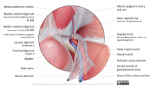
The base image is Male inguinal area internal view - no labels, illustration by Ron Slagter. Labels by Paul Gobée, MD. The image shows the inguinal area as viewed internally, for instance during laparoscopy. On the first slide the structures are labeled, on the second and third slide the entry points of inguinal and femoral hernia’s are labeled.
The base image is Male inguinal area internal view - no labels, illustration by Ron Slagter. Labels by Paul Gobée, MD. The image shows the inguinal area as viewed internally, for instance during laparoscopy. On the first slide the structures are labeled, on the second and third slide the entry points of inguinal and femoral hernia’s are labeled.
Anatomical structures in item:
Uploaded by: opgobee
Netherlands, Leiden – Leiden University Medical Center, Leiden University
Ductus deferens
Arteria testicularis
Vena testicularis
Ramus genitalis nervus genitofemoralis
Arteria iliaca externa
Vena iliaca externa
Arteria epigastrica inferior
Vena epigastrica inferior
Plica umbilicalis medialis
Plica umbilicalis mediana
Ligamentum umbilicale medianum
Ligamentum umbilicale mediale
Ramus pubicus ateria epigastricae inferioris
Tractus iliopubicus
Ligamentum lacunare
Ligamentum pectineum
Musculus iliacus
Musculus psoas major
Anulus inguinalis profundus
Anulus femoralis
Fossa inguinalis lateralis
Inguen
Creator(s)/credit: O. Paul Gobée MD, anatomist, LUMC; Ron Slagter NZIMBI, Medical illustrator
Requirements for usage
You are free to use this item if you follow the requirements of the license:  View license
View license If you use this item you should credit it as follows:
- For usage in print - copy and paste the line below:
- For digital usage (e.g. in PowerPoint, Impress, Word, Writer) - copy and paste the line below (optionally add the license icon):
"Leiden - Presentation slides Inguinal area, internal view, and entry points of inguinal and femoral hernia’s" at AnatomyTOOL.org by O. Paul Gobée, LUMC and Ron Slagter, license: Creative Commons Attribution-NonCommercial-ShareAlike


Comments