
nid: 59191
Additional formats:
None available
Description:
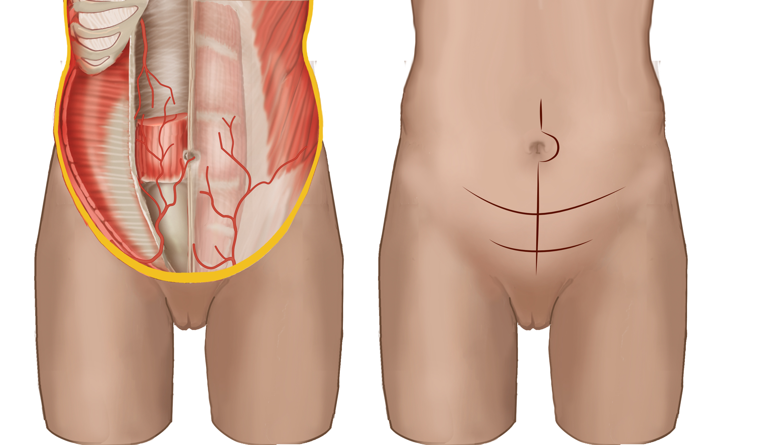
Different incisions for laparotomic approach pelvic surgery. On the left the anterior abdominal wall muscles and the inferior epigastric artery lying deep to the rectus muscle on the posterior rectus sheath, are shown. The superficial epigastric artery lies anterior to the anterior rectus sheath. To the right the Pfannenstiel, Mayard and median incision are depicted. No labels.
Illustration by Ron Slagter and Marco DeRuiter for course 'Surgical Anatomy of the lesser pelvis' by the 'Urologisch Opleidings Instituut', the Netherlands.
Illustration by Ron Slagter and Marco DeRuiter for course 'Surgical Anatomy of the lesser pelvis' by the 'Urologisch Opleidings Instituut', the Netherlands.
Anatomical structures in item:
Uploaded by: Siem Zethof
Netherlands, Leiden – Leiden University Medical Center, Leiden University
Arteria epigastrica inferior
Arteria epigastrica superficialis
Musculi abdominis
Vagina musculi recti abdominis
Musculus rectus abdominis
Creator(s)/credit: Ron Slagter NZIMBI, medical illustrator, LUMC; Prof. Marco DeRuiter PhD, anatomist, LUMC
Requirements for usage
You are free to use this item if you follow the requirements of the license:  View license
View license If you use this item you should credit it as follows:
- For usage in print - copy and paste the line below:
- For digital usage (e.g. in PowerPoint, Impress, Word, Writer) - copy and paste the line below (optionally add the license icon):
"Slagter - Different incisions for laparotomic approach pelvic surgery - no labels" at AnatomyTOOL.org by Ron Slagter, LUMC and Marco DeRuiter, LUMC, license: Creative Commons Attribution-NonCommercial-ShareAlike
"Slagter - Different incisions for laparotomic approach pelvic surgery - no labels" by Ron Slagter, LUMC and Marco DeRuiter, LUMC, license: CC BY-NC-SA


Comments