
nid: 59180
Additional formats:
None available
Description:
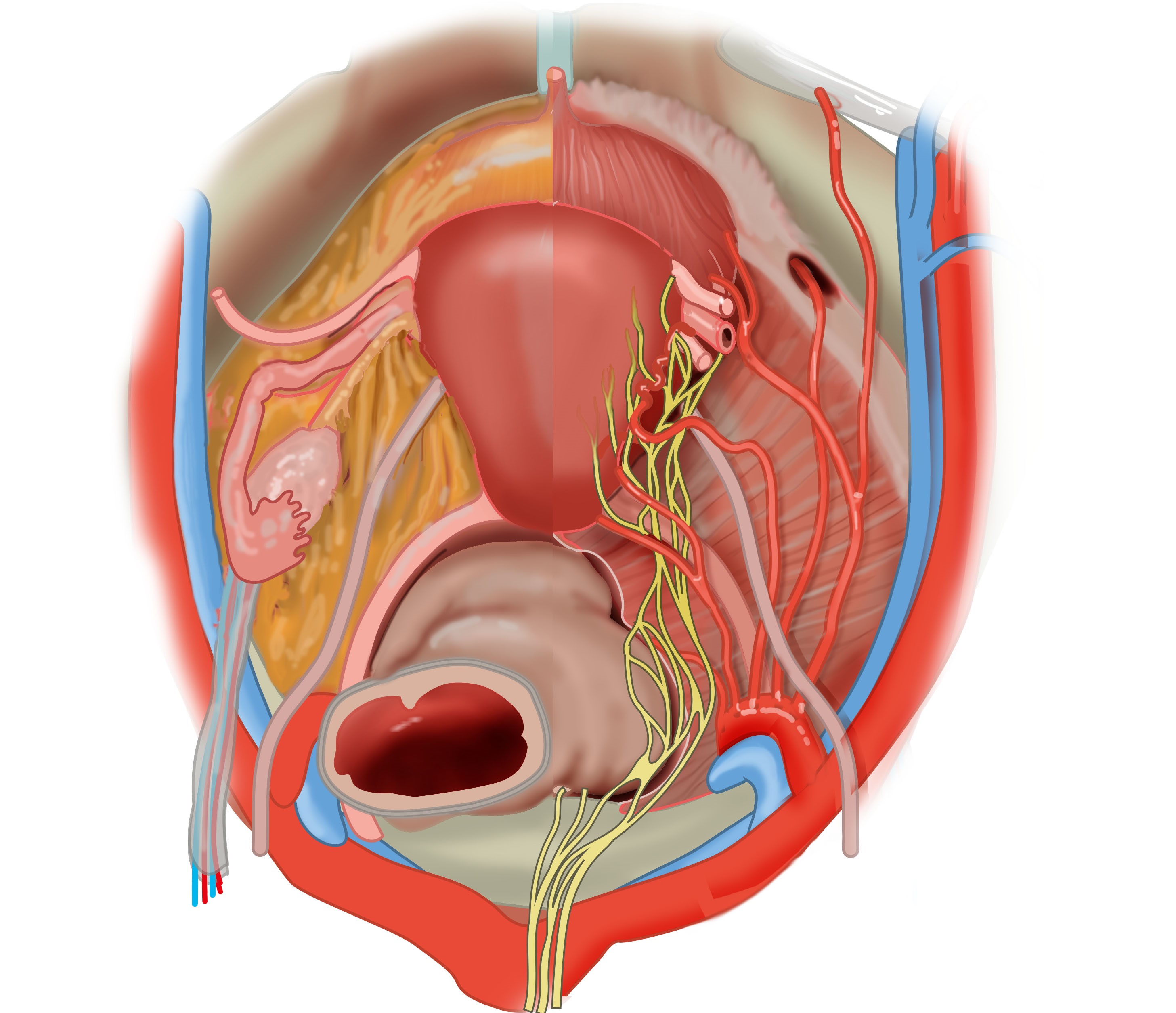
Superior view of the female pelvis without peritoneum. Left: the peritoneum has been removed. Structures shown on the lateral side of the uterus, from anterior to posterior: round ligament of the uterus, uterine tube, ovarian ligament, ureter, uterosacral ligament. The ovary is connected to the lateral pelvic wall with the suspensory ligament of the ovary. Right: the endopelvic fascia, ovary, uterine tube and round ligament have been removed to show the underlying arterial supply and pelvic plexus (inferior hypogastric plexus). The branches supplying the bladder, uterus and rectum originate from the internal iliac artery. The ureter crosses the externa iliac artery and runs medio-inferior going under the uterine artery before it enters the bladder ("water goes under the bridge"). The inferior hypogastric plexus (pelvic plexus) is located lateral to the rectum. The urachus lies anterosuperior on the bladder. No labels.
Illustration by Ron Slagter and Marco DeRuiter for course 'Surgical Anatomy of the lesser pelvis' by the 'Urologisch Opleidings Instituut', the Netherlands.
Illustration by Ron Slagter and Marco DeRuiter for course 'Surgical Anatomy of the lesser pelvis' by the 'Urologisch Opleidings Instituut', the Netherlands.
Anatomical structures in item:
Uploaded by: Siem Zethof
Netherlands, Leiden – Leiden University Medical Center, Leiden University
Ligamentum teres uteri
Tuba uterina (Salpinx)
Uterus
Ligamentum ovarii proprium
Ureter
Ligamentum rectouterinum
Ligamentum suspensorium ovarii
Arteria iliaca interna
Arteria obturatoria
Arteria uterina
Plexus nervosus hypogastricus inferior
Pelvis
Creator(s)/credit: Ron Slagter NZIMBI, medical illustrator, LUMC; Prof. Marco DeRuiter PhD, anatomist, LUMC
Requirements for usage
You are free to use this item if you follow the requirements of the license:  View license
View license If you use this item you should credit it as follows:
- For usage in print - copy and paste the line below:
- For digital usage (e.g. in PowerPoint, Impress, Word, Writer) - copy and paste the line below (optionally add the license icon):
"Superior view of the female pelvis with hypogastric plexus and internal iliac artery branches - no labels " at AnatomyTOOL.org by Ron Slagter, LUMC and Marco DeRuiter, LUMC, license: Creative Commons Attribution-NonCommercial-ShareAlike


Comments