
nid: 63195
Additional formats:
None available
Description:
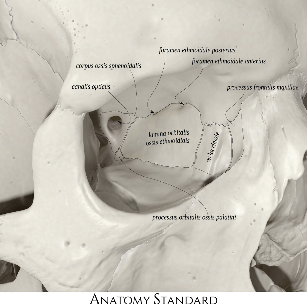
Medial wall of the orbit. The orbit is the skull's compartment containing the eyeball, extraocular muscles, lacrimal gland, ophthalmic blood vessels, and multiple cranial nerves. This image shows the medial wall. In terms of the skeleton, the medial wall of the orbit is the most complex one. Note the orbital process of palatine bone that forms a fraction of the wall, but is a useful anatomical landmark for endoscopic endonasal surgical approach to the orbit's medial wall. Latin labels.
The anatomy of the other three walls of the orbit can be found here.
Image and description retrieved from Anatomy Standard.
The anatomy of the other three walls of the orbit can be found here.
Image and description retrieved from Anatomy Standard.
Anatomical structures in item:
Uploaded by: rva
Netherlands, Leiden – Leiden University Medical Center, Leiden University
Orbita
Paries medialis orbitae
Corpus ossis sphenoidalis
Canalis opticus
Foramen ethmoidale posterius
Foramen ethmoidale anterius
Processus frontalis maxillae
Lamina orbitalis ossis ethmoidalis
Os lacrimale
Processus orbitalis ossis palatini
Creator(s)/credit: Jānis Šavlovskis MD, PhD, Assistant Professor; Kristaps Raits, 3D generalist
Requirements for usage
You are free to use this item if you follow the requirements of the license:  View license
View license If you use this item you should credit it as follows:
- For usage in print - copy and paste the line below:
- For digital usage (e.g. in PowerPoint, Impress, Word, Writer) - copy and paste the line below (optionally add the license icon):
"Anatomy Standard - Drawing Medial wall of the orbit - Latin labels" at AnatomyTOOL.org by Jānis Šavlovskis and Kristaps Raits, license: Creative Commons Attribution-NonCommercial


Comments