|
Data item |
Dundee - 3D model Regions of the brain |
|
rva |
Creative Commons Attribution-NonCommercial-ShareAlike |
3.402 |
9 |
|
Data item |
Slagter - Drawing Deep and superficial veins of the lower extremity - Dutch labels |
 |
rva |
Creative Commons Attribution-NonCommercial-ShareAlike |
3.395 |
2 |
|
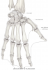
Data item |
OpenStax AnatPhys fig.8.7 - Hand and Wrist - English labels |
 |
Jorn IJkhout |
Creative Commons Attribution |
3.386 |
12 |
|
Data item |
OpenStax AnatPhys fig.6.12 - Diagram of Compact Bone - English labels |
 |
Jorn IJkhout |
Creative Commons Attribution |
3.384 |
6 |
|
Data item |
Leiden MOOC - Anatomy of the Abdomen and Pelvis; a journey from basis to clinic |
|
opgobee |
Creative Commons Attribution-NonCommercial-ShareAlike |
3.366 |
3 |
|
Data item |
Coronal section of the female pelvis showing the cervix, vagina, cardinal ligament and levator ani muscle – Dutch labels |
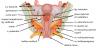 |
Siem Zethof |
Creative Commons Attribution-NonCommercial-ShareAlike |
3.366 |
1 |
|
Learning Path |
Quiz Aorta Abdominalis Takken (basis) |
|
tjscherphof |
Creative Commons Attribution-NonCommercial-ShareAlike |
3.360 |
3 |
|
Data item |
Anatomy Standard - Drawing Atlanto-occipital and atlanto-axial joints: anterior view - Latin labels |
 |
rva |
Creative Commons Attribution-NonCommercial |
3.359 |
11 |
|
Data item |
Slagter - Drawing Insertion of thumb muscles - Dutch labels |
 |
rva |
Creative Commons Attribution-NonCommercial-ShareAlike |
3.353 |
10 |
|
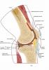
Data item |
Anatomy Standard - Drawing Anatomy of the distal femur - Latin labels |
 |
rva |
Creative Commons Attribution-NonCommercial |
3.332 |
4 |
|
Anatomical Structure |
Ala major ossis sphenoidalis |
|
admin |
|
3.317 |
4 |
|
Data item |
Anatomy Standard - Drawing Anterior and posterior view of lower leg bones - no labels |
 |
rva |
Creative Commons Attribution-NonCommercial |
3.281 |
15 |
|
Data item |
Minnesota Atlas Card Anat - Video Beating Mitral Valve |
|
opgobee |
This item is (on) an external site. The license as stated on that site holds. |
3.279 |
7 |
|
Data item |
Groningen - 3D model Heart with Right Dominance - numbered English and Latin labels |
|
rva |
Creative Commons Attribution-NonCommercial-ShareAlike |
3.266 |
5 |
|
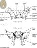
Data item |
Anterior view of female pelvis; internal organs and innervation - Latin and English labels |
 |
admin |
Creative Commons Attribution-NonCommercial-ShareAlike |
3.257 |
4 |
|
Data item |
Leiden - Schema Autonome Zenuwstelsel - Dutch, Latin labels |
|
opgobee |
Creative Commons Attribution-NonCommercial-ShareAlike |
3.255 |
3 |
|
Data item |
NAV Congress 2022 - Poster AnatomyTOOL survey Students 2021: results and measures taken |
|
opgobee |
Creative Commons Attribution-ShareAlike |
3.248 |
6 |
|
Data item |
U.Br.Columbia - Drawing Caudate nucleus and amygdala - English labels |
 |
rva |
Creative Commons Attribution-NonCommercial-ShareAlike |
3.245 |
7 |
|
Data item |
Leiden - Video Cattell-Braasch manoeuvre, unhiding the duodenum |
|
opgobee |
Creative Commons Attribution-NonCommercial-ShareAlike |
3.244 |
4 |
|
Learning Path |
Quiz Large Intestine - Characteristics and Parts (basics) |
|
EmmaL |
Creative Commons Attribution-NonCommercial-ShareAlike |
3.244 |
0 |
|
Data item |
NYSORA - Drawing Intermediate muscles of back (erector spinae) - English labels |
 |
rva |
Creative Commons Attribution-NonCommercial-NoDerivs |
3.243 |
3 |
|
Data item |
KnowledgeWorks - Drawing Abdominal organs - no labels |
 |
rva |
Creative Commons Attribution-NonCommercial-ShareAlike |
3.241 |
8 |
|
Data item |
Dundee - 3D model Cranial Nerves |
|
rva |
Creative Commons Attribution-ShareAlike |
3.236 |
1 |
|
Data item |
Nervus fibularis superficialis |
 |
anatomiekulak |
Creative Commons Attribution-NonCommercial |
3.233 |
5 |
|
Data item |
Elon - 3D model Fibula |
|
rva |
Creative Commons Attribution |
3.231 |
7 |
|
Data item |
NAV Congress 2022 - Lunch workshop AnatomyTOOL quizzen maken |
|
opgobee |
Creative Commons Attribution-ShareAlike |
3.223 |
7 |
|
Data item |
NAV Congress 2022 - Poster AnatomyTOOL survey Anatomists 2021: results and measures taken |
|
opgobee |
Creative Commons Attribution-ShareAlike |
3.205 |
5 |
|
Learning Path |
Quiz Buikwand - Kenmerken en Onderdelen (basis) |
|
tjscherphof |
Creative Commons Attribution-NonCommercial-ShareAlike |
3.204 |
2 |
|
Data item |
Elon - 3D model Calcaneus |
|
rva |
Creative Commons Attribution |
3.201 |
5 |
|
Data item |
Anatomy Standard - Drawing Frontal bone: anterior view - Latin labels |
 |
rva |
Creative Commons Attribution-NonCommercial |
3.190 |
3 |
|
Learning Path |
LUMC G1St Ingangstoets Practicum |
|
opgobee |
Creative Commons Attribution-NonCommercial-ShareAlike |
3.179 |
1 |
|
Data item |
Minnesota Atlas Card Anat - Video Beating Aortic and Mitral Valve |
|
opgobee |
This item is (on) an external site. The license as stated on that site holds. |
3.179 |
7 |
|
Data item |
Elon - 3D model Humerus |
|
rva |
Creative Commons Attribution |
3.175 |
18 |
|
Data item |
RCSI - Drawing Internal surface of cranial base - English labels |
 |
rva |
Creative Commons Attribution-NonCommercial-ShareAlike |
3.163 |
10 |
|
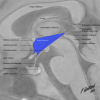
Data item |
Rotation video of 3D reconstruction female pelvis, pelvic diaphragm and organs - build, English labels |
|
opgobee |
Creative Commons Attribution-NonCommercial-ShareAlike |
3.159 |
6 |
|
Data item |
OpenStax AnatPhys fig.19.10 - Congenital Heart Defects - English labels |
 |
Jorn IJkhout |
Creative Commons Attribution |
3.155 |
2 |
|
Data item |
Leiden - Video Demonstratie Anatomie Abdomen Deel 2 (dissectie-preparaat) |
|
rva |
Unknown license |
3.150 |
2 |
|
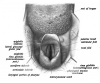
Data item |
Rectus abdominis muscle |
|
opgobee |
Creative Commons Attribution-NonCommercial-ShareAlike |
3.149 |
4 |
|
Anatomical Structure |
Condylus humeri |
|
admin |
|
3.146 |
7 |
|
Data item |
Neuroanatomy - collection page |
|
PM |
Creative Commons Attribution-NonCommercial-ShareAlike |
3.133 |
7 |
|
Data item |
Investigation Anatomy Learning Resources - which are open? |
|
lumctest |
Creative Commons Attribution-ShareAlike |
3.113 |
2 |
|
Data item |
OpenStax AnatPhys fig.7.10 - Sphenoid Bone - English labels |
 |
Jorn IJkhout |
Creative Commons Attribution |
3.112 |
3 |
|
Data item |
Radiopaedia - Drawing Boundaries of the hypothalamus with translucent MRI - English labels |
 |
rva |
Creative Commons Attribution-NonCommercial-ShareAlike |
3.109 |
6 |
|
Data item |
RCSI - Drawing Knee joint: sagittal section - English labels |
 |
rva |
Creative Commons Attribution-NonCommercial-ShareAlike |
3.106 |
7 |
|
Data item |
Sobotta 1906 fig.438 - Aditus laryngis - English labels |
 |
Student128 |
Public Domain |
3.089 |
4 |
|
Data item |
Prostate nerve and venous plexus, innervation and venous drainage penis – No labels |
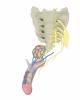 |
Siem Zethof |
Creative Commons Attribution-NonCommercial-ShareAlike |
3.086 |
8 |
|
Data item |
Slagter - Drawing Male inguinal area internal view - English labels |
 |
opgobee |
Creative Commons Attribution-NonCommercial-ShareAlike |
3.069 |
3 |
|
Data item |
Dundee - Drawing Posterior and Anterior View of the Larynx - English labels |
 |
rva |
Creative Commons Attribution-ShareAlike |
3.061 |
7 |
|
Data item |
BlueLink - 3D model Fifth Cervical Vertebra (C5) |
|
rva |
Creative Commons Attribution-NonCommercial-NoDerivs |
3.053 |
6 |
|
Anatomical Structure |
Facies auricularis ossis sacri |
|
admin |
|
3.051 |
5 |
|
Data item |
colon x-ray contrast |
 |
aherrler |
Creative Commons Attribution-ShareAlike |
3.042 |
3 |
|
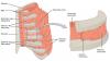
Data item |
Human Biology fig. 1.20 - Structure of a skeletal muscle - English labels |
 |
rva |
Public Domain |
3.030 |
7 |
|
Data item |
Internal abdominal oblique muscle |
|
opgobee |
Creative Commons Attribution-NonCommercial-ShareAlike |
3.007 |
3 |
|
Anatomical Structure |
Humor aquosus |
|
admin |
|
2.987 |
5 |
|
Learning Path |
BBS1002 Gastrointestinal tract Anatomy Self-study |
|
leo.koehler |
Creative Commons Attribution-NonCommercial-ShareAlike |
2.974 |
0 |
|
Data item |
Jain - Drawing Anterior view of heart - no labels |
 |
rva |
Creative Commons Attribution-NonCommercial-NoDerivs |
2.967 |
4 |
|
Anatomical Structure |
Margo medialis scapulae |
|
admin |
|
2.965 |
5 |
|
Data item |
About Medicine: 3D model of anterior forearm muscles |
|
opgobee |
Creative Commons Attribution |
2.946 |
5 |
|
Data item |
OpenStax AnatPhys fig.11.18 - Thorax - English labels |
 |
Jorn IJkhout |
Creative Commons Attribution |
2.944 |
4 |
|
Data item |
Photo Intravaginal view of normal cervix uteri - Latin and English labels |
 |
opgobee |
Creative Commons Attribution-ShareAlike |
2.915 |
5 |
|
Data item |
Minnesota Atlas Card Anat - Video Beating Tricuspid Valve |
|
opgobee |
This item is (on) an external site. The license as stated on that site holds. |
2.891 |
5 |
|
Data item |
Dundee - Drawing The superficial lymph nodes of the head and neck - English labels |
 |
rva |
Creative Commons Attribution-NonCommercial-NoDerivs |
2.884 |
2 |
|
Anatomical Structure |
Recessus costodiaphragmaticus |
|
admin |
|
2.878 |
10 |
|
Data item |
OpenStax AnatPhys fig.12.27 - Chemical Synapse - English labels |
 |
Jorn IJkhout |
Creative Commons Attribution |
2.873 |
7 |
|
Data item |
Presentatie Waarom Open Onderwijs Materialen gebruiken (kort) |
|
opgobee |
Creative Commons Attribution-ShareAlike |
2.866 |
3 |
|
Data item |
KnowledgeWorks - Drawing Overview of endocrine glands - English labels |
 |
rva |
Creative Commons Attribution |
2.865 |
9 |
|
Data item |
LadyofHats - Drawing Branches of the aorta - English labels |
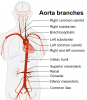 |
rva |
Creative Commons Attribution-ShareAlike |
2.860 |
4 |
|
Data item |
OpenStax AnatPhys fig.4.10 - Modes of Secretion by Glands updated - English labels |
 |
Jorn IJkhout |
Creative Commons Attribution |
2.858 |
8 |
|
Data item |
Cenveo - Drawing Liver anatomy and vascularisation - English labels |
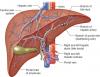 |
rva |
Creative Commons Attribution |
2.858 |
7 |
|
Data item |
Lateral view of opened thorax with right lung |
 |
Nadja Baltensweiler |
Creative Commons Attribution-NonCommercial-ShareAlike |
2.857 |
6 |
|
Data item |
Anatomy Standard - Drawing Thorcacic vertebra (Th4): superior view - Latin labels |
 |
rva |
Creative Commons Attribution-NonCommercial |
2.856 |
9 |
|
Data item |
Lecturio - Drawing Cross-section of the arm and its compartments - English labels |
 |
rva |
Creative Commons Attribution-NonCommercial-ShareAlike |
2.856 |
8 |
|
Data item |
Presentation Why use Open Educational Resources (OER)? |
|
opgobee |
Creative Commons Attribution-ShareAlike |
2.851 |
5 |
|
Data item |
Human Biology fig. 1.106 - Blastocyst before implantation - English labels |
 |
rva |
Creative Commons Attribution-NonCommercial |
2.848 |
3 |
|
Learning Path |
Quiz Heart - Valves (advanced) |
|
EmmaL |
Creative Commons Attribution-NonCommercial-ShareAlike |
2.844 |
3 |
|
Data item |
Minnesota Atlas Card Anat - Video Beating Pulmonary Valve |
|
opgobee |
This item is (on) an external site. The license as stated on that site holds. |
2.844 |
5 |
|
Data item |
Leiden - Drawing Overview Digestive Tract - no labels |
 |
opgobee |
Creative Commons Attribution-NonCommercial-ShareAlike |
2.827 |
8 |
|
Data item |
Blausen 0723 - Anatomy of the pelvis - English labels |
 |
Student10 |
Creative Commons Attribution |
2.806 |
1 |
|
Data item |
Elon - 3D model Skull of the Fetus |
|
rva |
Creative Commons Attribution |
2.803 |
12 |
|
Data item |
Anatomy Standard - Drawing Temporal bone: posterior view - Latin labels |
 |
rva |
Creative Commons Attribution-NonCommercial |
2.802 |
2 |
|
Data item |
Dundee - Drawing Anatomy of the ear - No labels |
 |
rva |
Creative Commons Attribution-NonCommercial-NoDerivs |
2.799 |
8 |
|
Anatomical Structure |
Ostium pharyngeum tubae auditivae |
|
admin |
|
2.799 |
4 |
|
Data item |
Dorsal foot nerves |
 |
anatomiekulak |
Creative Commons Attribution-NonCommercial |
2.794 |
5 |
|
Data item |
Lexington - 3D model Thoracic vertebra |
|
rva |
Creative Commons Attribution |
2.794 |
1 |
|
Data item |
Anatomy Standard - Drawing Bones of hand: dorsal view - Latin labels |
 |
rva |
Creative Commons Attribution-NonCommercial |
2.793 |
8 |
|
Data item |
External abdominal oblique muscle |
|
opgobee |
Creative Commons Attribution-NonCommercial-ShareAlike |
2.788 |
4 |
|
Data item |
Dundee - Drawing Anatomy of the Mastoid Antrum - English labels |
 |
rva |
Creative Commons Attribution-NonCommercial-NoDerivs |
2.780 |
4 |
|
Data item |
Radiopaedia - Drawing Anatomy of the brain ventricles - English labels |
 |
rva |
Creative Commons Attribution-NonCommercial-NoDerivs |
2.767 |
6 |
|
Simple page |
Open 3D Model |
|
admin |
|
2.759 |
61 |
|
Data item |
Leiden - Drawing Entry points of inguinal and femoral hernia's, internal view - English labels |
 |
opgobee |
Creative Commons Attribution-NonCommercial-ShareAlike |
2.750 |
3 |
|
Data item |
Normal heart |
|
kmeier |
Creative Commons Attribution-NonCommercial-ShareAlike |
2.749 |
2 |
|
Data item |
Anatomy Standard - Drawing Talus bone ex situ: superior posterior view - Latin labels |
 |
rva |
Creative Commons Attribution-NonCommercial |
2.748 |
7 |
|
Data item |
LadyofHats - Drawing Lateral skull anatomy - Latin labels |
 |
rva |
Public Domain |
2.747 |
4 |
|
Learning Path |
Quiz Maag - Vascularisatie (basis) |
|
tjscherphof |
Creative Commons Attribution-NonCommercial-ShareAlike |
2.743 |
0 |
|
Data item |
Servier - Drawing Layers of vein and artery - English labels |
 |
rva |
Creative Commons Attribution |
2.741 |
5 |
|
Data item |
Cross-sectional view of the penis with layered structure visible - Dutch labels |
 |
Siem Zethof |
Creative Commons Attribution-NonCommercial-ShareAlike |
2.736 |
18 |
|
Data item |
Fundus cavitas oris, facies superioris |
 |
koolstra |
Creative Commons Attribution-NonCommercial-ShareAlike |
2.722 |
3 |
|
Data item |
Radiopaedia - Drawing Aortic arch and its branches - English labels |
 |
rva |
Creative Commons Attribution-NonCommercial-NoDerivs |
2.714 |
7 |
|
Learning Path |
Quiz Biliary tract (basics) |
|
EmmaL |
Creative Commons Attribution-NonCommercial-ShareAlike |
2.706 |
5 |
|
Data item |
Leiden, Maas Photo 30 - Female pelvis view from anterior (plastination specimen) - number labels with legend |
 |
opgobee |
Creative Commons Attribution-NonCommercial-ShareAlike |
2.704 |
2 |