
nid: 58133
Additional formats:
None available
Description:
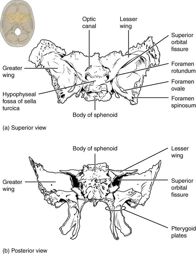
Sphenoid Bone. Shown in isolation in (a) superior and (b) posterior views, the sphenoid bone is a single midline bone that forms the anterior walls and floor of the middle cranial fossa. It has a pair of lesser wings and a pair of greater wings. The sella turcica surrounds the hypophyseal fossa. Projecting downward are the medial and lateral pterygoid plates. The sphenoid has multiple openings for the passage of nerves and blood vessels, including the optic canal, superior orbital fissure, foramen rotundum, foramen ovale, and foramen spinosum. English labels. From OpenStax book 'Anatomy and Physiology', fig. 7.10.
Anatomical structures in item:
Uploaded by: Jorn IJkhout
Netherlands, Leiden – Leiden University Medical Center, Leiden University
Bone
Os sphenoidale
Corpus ossis sphenoidalis
Ala major ossis sphenoidalis
Ala minor ossis sphenoidalis
Processus pterygoideus ossis sphenoidalis
Fossa hypophysialis
Creator(s)/credit: OpenStax
Requirements for usage
You are free to use this item if you follow the requirements of the license:  View license
View license If you use this item you should credit it as follows:
- For usage in print - copy and paste the line below:
- For digital usage (e.g. in PowerPoint, Impress, Word, Writer) - copy and paste the line below (optionally add the license icon):
"OpenStax AnatPhys fig.7.10 - Sphenoid Bone - English labels" at AnatomyTOOL.org by OpenStax, license: Creative Commons Attribution. Source: book 'Anatomy and Physiology', https://openstax.org/details/books/anatomy-and-physiology.
"OpenStax AnatPhys fig.7.10 - Sphenoid Bone - English labels" by OpenStax, license: CC BY. Source: book 'Anatomy and Physiology', https://openstax.org/details/books/anatomy-and-physiology.


Comments