
nid: 60841
Additional formats:
None available
Description:
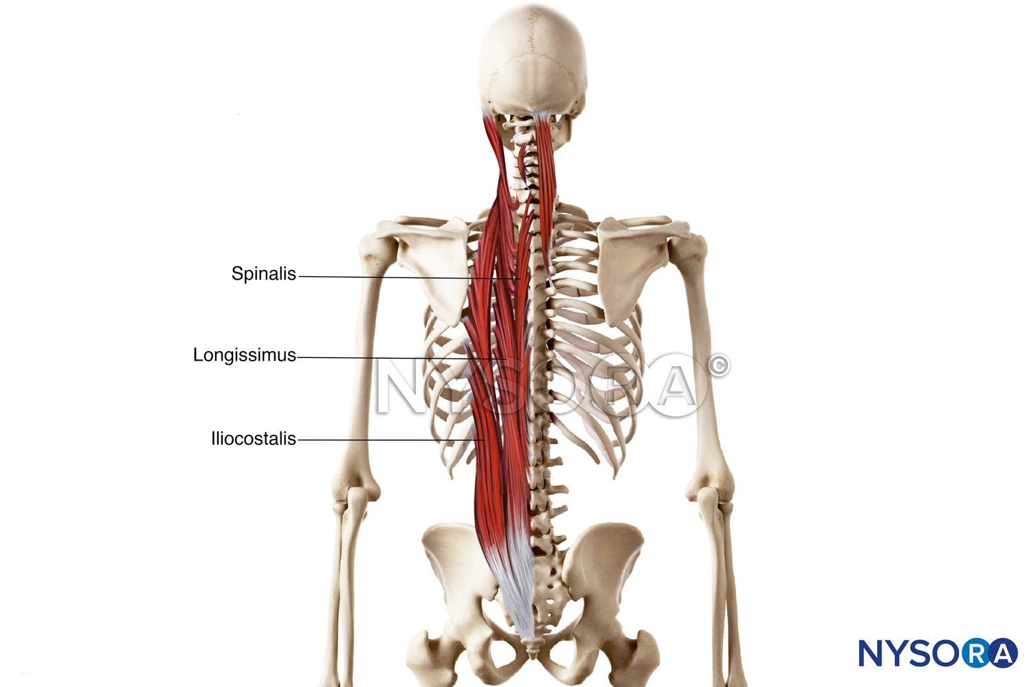
Intermediate muscles of back (erector spinae). The anatomy of the intermediate muscles of the back can be seen. The spinalis, longissiumus and iliocostalis muscle form a column known as the erector spinae. Also some other muscles are shown but not labeled. English labels.
Image created for NYSORA by VisionExpo.Design - www.nysora.com
Image created for NYSORA by VisionExpo.Design - www.nysora.com
Anatomical structures in item:
Uploaded by: rva
Netherlands, Leiden – Leiden University Medical Center, Leiden University
Musculus spinalis
Musculus erector spinae
Musculus longissimus
Musculus iliocostalis
Dorsum
Creator(s)/credit: New York School of Regional Anesthesia; VisionExpo.Design, illustration creation
Requirements for usage
You are free to use this item if you follow the requirements of the license:  View license
View license If you use this item you should credit it as follows:
- For usage in print - copy and paste the line below:
- For digital usage (e.g. in PowerPoint, Impress, Word, Writer) - copy and paste the line below (optionally add the license icon):
"NYSORA - Drawing Intermediate muscles of back (erector spinae) - English labels" at AnatomyTOOL.org by New York School of Regional Anesthesia and VisionExpo.Design, license: Creative Commons Attribution-NonCommercial-NoDerivs


Comments