
nid: 62719
Additional formats:
None available
Description:
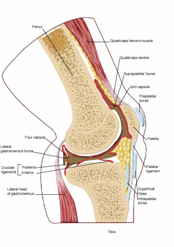
Knee joint: sagittal section. This drawing shows the bones, muscles, bursae, and ligaments of the knee. English labels.
This image by the Royal College of Surgeons of Ireland (RCSI) is retrieved from Health Education Assets Library (HEAL) of the University of Utah.
This image by the Royal College of Surgeons of Ireland (RCSI) is retrieved from Health Education Assets Library (HEAL) of the University of Utah.
Anatomical structures in item:
Uploaded by: rva
Netherlands, Leiden – Leiden University Medical Center, Leiden University
Articulatio genus
Femur
Musculus quadriceps femoris
Bursa suprapatellaris
Capsula articularis
Ligamentum patellae
Patella
Tibia
Bursa infrapatellaris profunda
Corpus adiposum infrapatellare
Bursa subcutanea infrapatellaris
Caput laterale musculus gastrocnemii
Ligamentum cruciatum anterius
Ligamentum cruciatum posterius
Creator(s)/credit: Royal College of Surgeons of Ireland
Requirements for usage
You are free to use this item if you follow the requirements of the license:  View license
View license If you use this item you should credit it as follows:
- For usage in print - copy and paste the line below:
- For digital usage (e.g. in PowerPoint, Impress, Word, Writer) - copy and paste the line below (optionally add the license icon):
"RCSI - Drawing Knee joint: sagittal section - English labels" at AnatomyTOOL.org by Royal College of Surgeons of Ireland, license: Creative Commons Attribution-NonCommercial-ShareAlike


Comments