Skull
A lateral view depicting the bones forming the cranium.
A superior view of the cranial floor and several its foramina.
Meninges
This video reviews the meninges and relevant pathology.
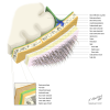
A schematic image illustrating the skull and meninges.
Views / cortex
This video demonstrates the cortex regions and somatic representation.
A ventral view of a dissected brain, showing its cortex and several neural and vascular structures.
A ventral view visualizing the cerebral cortex (including gyri and sulci) and a section of the midbrain.
A midsagittal view of a dissected brain, depicting its cortex and vascular structures.
A midsagittal view, visualizing the cortex including various gyri and sulci.
A dorsal view of a dissected brain, showing a largely intact arachnoid; a portion of the brain has been cut to show white matter.
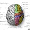
A dorsal view of the brain, visualizing its cortex including various gyri and sulci.
A lateral view of the brain, depicting its cortex including various gyri and sulci.
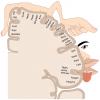
A schematic image visualizing the organization of the sensory homunculus.
Ventricles
This video demonstrates the ventricular system.
A schematic image visualizing the ventricle system.
Basal ganglia
This video covers the basal ganglia and several relevant pathways.
A lateral view of the brain depicting several basal ganglia.
A horizontal section of the brain visualizing various basal ganglia and its relation to the ventricles.
This video covers the basal ganglia and several relevant pathways.
A lateral view of the brain visualizing the limbic system and its components.
Cranial nerves
A video covering the cranial nerves on a specimen.
A ventral view of the brain depicting the twelve cranial nerves.
A dorsal view of the cranial base, visualizing the cranial nerves entering their respective foramina.
This video covers three critical vertical pathways.
A schematic image illustrating the optic tract.
A schematic overview of several fiber tracts and its locations in the brain and spinal cord.
Vascularisation - Arteries
This video discusses the cerebral circulation.
A midsagittal view of the brain visualizing various arteries.
A ventral view of the brain depicting the circle of Willis.
A schematic lateral view of the brain illustrating the areas vasculated by the three main cerebral arteries.
A schematic midsagittal view of the brain illustrating the areas vasculated by the three main cerebral arteries.
Vascularisation - Veins
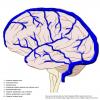
A schematic lateral view of the brain depicting its venous structures.
A schematic illustration visualizing the sinuses of the brain.
Spinal Cord
This video highlights the different aspects of the spinal cord.
This video discusses the organization of the nervous system.
A schematic image illustrating the connections of the parasympathetic nervous system.
A schematic image illustrating the connections of the (ortho)sympathetic nervous system.
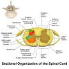
A horizontal view of the spinal cord visualizing its organisation of nervous structures.
A schematic illustration of the spinal cord and its respective roots.
Overview
A schematic overview of the largest nervous structures.
A schematic overview of the dermatomes and its respective nerves.
A schematic image illustrating the brachial plexus and its relevant nerves.
More
Many more great open neuroanatomical resources are present on the Website NeuroAnatomy of the Univ. of Br. Columbia. Find the reference on the page 'Best Open Anatomy Learning Resources'.
Many great open neuroanatomical tutorial dissection videos were created by prof Suzanne Stensaas of the Univ. of Utah and distributed as 'Neuroanatomy Video lab'. Find the reference on the page 'Best Open Anatomy Learning Resources'.
Cross-sections
 View license
 If you use this item you should credit it as follows:
- For usage in print - copy and paste the line below:
- For digital usage (e.g. in PowerPoint, Impress, Word, Writer) - copy and paste the line below (optionally add the license icon):
"Neuroanatomy - collection page" by Alexander R. J. Bijnsdorp, VUMC, license: CC BY-NC-SA











Comments