

nid: 62540
Additional formats:
None available
Description:
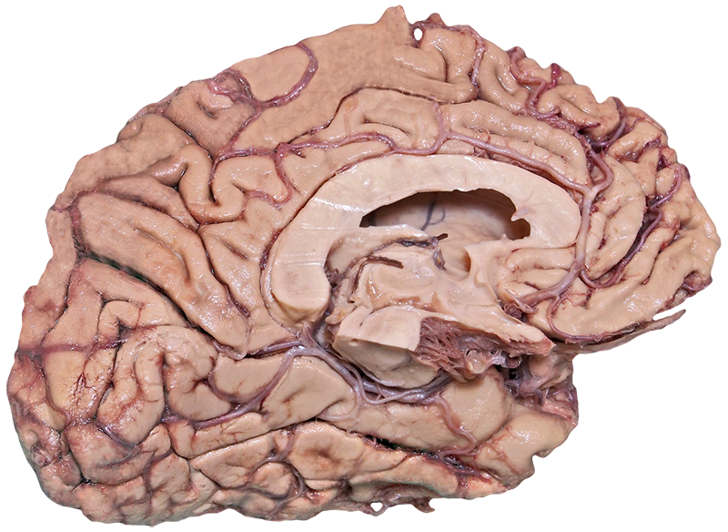
Cortex: medial view. In this figure, a dissection of the brain is shown. The specimen shows the corpus callosum, fornix, hypothalamus, lateral ventricle, thalamus and several blood vessels of the brain. These structures can be made visible on this website. No labels.
Anatomical structures in item:
Uploaded by: rva
Netherlands, Leiden – Leiden University Medical Center, Leiden University
Arteria cerebri anterior
Truncus encephali
Corpus callosum
Fornix
Hypothalamus
Arteria pericallosa
Arteria cerebri posterior
Thalamus
Creator(s)/credit: Prof. Claudia Krebs MD, PhD, anatomist, UBC; Monika Fejtek, digital media technologist, UBC
Requirements for usage
You are free to use this item if you follow the requirements of the license:  View license
View license If you use this item you should credit it as follows:
- For usage in print - copy and paste the line below:
- For digital usage (e.g. in PowerPoint, Impress, Word, Writer) - copy and paste the line below (optionally add the license icon):
"U.Br.Columbia - Photo Cortex: medial view (dissection) - no labels" at AnatomyTOOL.org by Claudia Krebs, UBC and Monika Fejtek, UBC, license: Creative Commons Attribution-NonCommercial-ShareAlike
"U.Br.Columbia - Photo Cortex: medial view (dissection) - no labels" by Claudia Krebs, UBC and Monika Fejtek, UBC, license: CC BY-NC-SA


Comments