|
Data item |
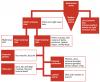
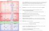
OpenStax AnatPhys fig.20.30 - Iliac Artery Branches Chart - English labels |
 |
Jorn IJkhout |
Creative Commons Attribution |
2.697 |
6 |
|
Data item |
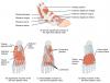
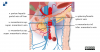
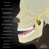
Radiopaedia - Drawing Main branches of the maxillary nerve - English labels |
 |
rva |
Creative Commons Attribution-NonCommercial-ShareAlike |
2.697 |
3 |
|
Data item |
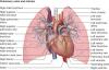
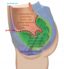
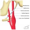
Lateral view of opened thorax, lung removed |
 |
Nadja Baltensweiler |
Creative Commons Attribution-NonCommercial-ShareAlike |
2.697 |
3 |
|
Data item |
Elon - 3D model Lumbar Vertebra |
|
rva |
Creative Commons Attribution-NonCommercial-NoDerivs |
2.691 |
9 |
|
Data item |
3D Heart models on NIH 3D print exchange website |
|
opgobee |
This item is (on) an external site. The license as stated on that site holds. |
2.689 |
5 |
|
Data item |
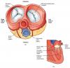
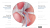
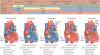
OpenStax AnatPhys fig.19.14 - Four Valves view during Systole - English labels |
 |
Jorn IJkhout |
Creative Commons Attribution |
2.681 |
4 |
|
Data item |
Drawing veins of stomach |
 |
lumcanatomy |
Creative Commons Attribution-NonCommercial-ShareAlike |
2.667 |
1 |
|
Data item |
OpenStax AnatPhys fig.20.42 - Lower Limb Veins Chart - English labels |
 |
Jorn IJkhout |
Creative Commons Attribution |
2.667 |
7 |
|
Data item |
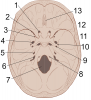
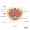
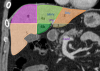
Leiden - Photo set Anatomy Cross-sections, caudal view |
|
opgobee |
Creative Commons Attribution-NonCommercial-ShareAlike |
2.663 |
3 |
|
Data item |
Dundee - 3D model Ligaments of the Female Pelvis |
|
rva |
Creative Commons Attribution-NonCommercial-ShareAlike |
2.657 |
4 |
|
Data item |
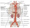
OpenStax AnatPhys fig.20.28 - Thoracic Abdominal Arteries - English labels |
 |
Jorn IJkhout |
Creative Commons Attribution |
2.635 |
12 |
|
Data item |
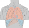
Lynch - Drawing Anterior chest landmarks - no labels |
 |
rva |
Creative Commons Attribution |
2.631 |
3 |
|
Data item |
Leiden - Drawing Entry points of inguinal and femoral hernia's, internal view - English labels |
 |
opgobee |
Creative Commons Attribution-NonCommercial-ShareAlike |
2.628 |
3 |
|
Data item |
OpenStax AnatPhys fig.11.34 - Intrinsic Muscles of the Foot - English labels |
 |
Jorn IJkhout |
Creative Commons Attribution |
2.625 |
13 |
|
Data item |
Elon - 3D model Axis (vertebra C2) |
|
rva |
Creative Commons Attribution |
2.608 |
7 |
|
Data item |
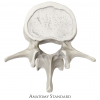
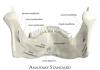
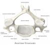
Anatomy Standard - Drawing Lumbar vertebra: superior view - no labels |
 |
rva |
Creative Commons Attribution-NonCommercial |
2.606 |
9 |
|
Anatomical Structure |
Incisura vertebralis superior |
|
admin |
|
2.599 |
8 |
|
Data item |
U.Br.Columbia - Drawing Posterior view of brainstem - English labels |
 |
rva |
Creative Commons Attribution-NonCommercial-ShareAlike |
2.598 |
12 |
|
Data item |
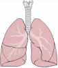
Lynch - Drawing Simplified anatomy of the lungs - no labels |
 |
rva |
Creative Commons Attribution |
2.594 |
10 |
|
Learning Path |
Quiz Lever - Kenmerken en Onderdelen (basis) |
|
tjscherphof |
Creative Commons Attribution-NonCommercial-ShareAlike |
2.589 |
2 |
|
Anatomical Structure |
Crista musculi supinatoris |
|
admin |
|
2.589 |
3 |
|
Data item |
OLI - Drawing Pulmonary bronchi, veins and arteries - English labels |
 |
rva |
Creative Commons Attribution-NonCommercial-ShareAlike |
2.584 |
2 |
|
Anatomical Structure |
Facies pelvica ossis sacri |
|
admin |
|
2.573 |
3 |
|
Anatomical Structure |
Massa lateralis atlantis |
|
admin |
|
2.562 |
5 |
|
Data item |
Leiden - Drawing Parts of the stomach - English labels |
 |
opgobee |
Creative Commons Attribution-NonCommercial-ShareAlike |
2.561 |
3 |
|
Data item |
Anatomy Standard - Drawing Atlas: inferior aspect - Latin labels |
 |
rva |
Creative Commons Attribution-NonCommercial |
2.555 |
3 |
|
Data item |
Tectum cavitas oris |
 |
koolstra |
Creative Commons Attribution-NonCommercial-ShareAlike |
2.549 |
5 |
|
Data item |
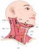
Slagter - Drawing Lymph node levels of the neck - labels |
 |
rva |
Creative Commons Attribution-NonCommercial-ShareAlike |
2.536 |
4 |
|
Data item |
Lynch - Drawing Internal surface of cranial base - numbered labels |
 |
rva |
Creative Commons Attribution |
2.529 |
6 |
|
Data item |
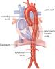
OLI - Drawing Parts of aorta in relation to heart and diaphragm - English labels |
 |
rva |
Creative Commons Attribution-NonCommercial-ShareAlike |
2.527 |
6 |
|
Data item |
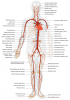
LadyofHats - Drawing Overview of the large arteries - English labels |
 |
rva |
Public Domain |
2.525 |
8 |
|
Data item |
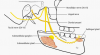
Lingua, systema vasculare |
 |
koolstra |
Creative Commons Attribution-NonCommercial-ShareAlike |
2.523 |
3 |
|
Data item |
RCSI - Drawing Flexor muscles and tendons of forearm - English labels |
 |
rva |
Creative Commons Attribution-NonCommercial-ShareAlike |
2.510 |
9 |
|
Anatomical Structure |
Fornix pharyngis |
|
admin |
|
2.500 |
3 |
|
Data item |
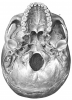
Sobotta 1909 fig.41 - The skull, inferior view - No labels |
 |
Student128 |
Public Domain |
2.499 |
3 |
|
Data item |
Abdominal wall (transverse section) |
 |
lumcanatomy |
Creative Commons Attribution-NonCommercial-ShareAlike |
2.498 |
9 |
|
Data item |
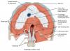
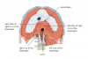
Slagter - Drawing Inferior view of the female pelvic diaphragm 2 - English labels |
 |
opgobee |
Creative Commons Attribution-NonCommercial-ShareAlike |
2.487 |
2 |
|
Data item |
Jmarchn - Drawing Human Anus - English labels |
 |
opgobee |
Creative Commons Attribution-ShareAlike |
2.484 |
6 |
|
Data item |
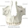
Anatomy Standard - Drawing Maxilla: anterior view - Latin labels |
 |
rva |
Creative Commons Attribution-NonCommercial |
2.482 |
5 |
|
Learning Path |
Quiz Inwendige Genitaliën Vrouw - Ligamenten (basis) |
|
tjscherphof |
Creative Commons Attribution-NonCommercial-ShareAlike |
2.482 |
3 |
|
Learning Path |
Quiz Dunne Darm - Kenmerken en Onderdelen (basis) |
|
tjscherphof |
Creative Commons Attribution-NonCommercial-ShareAlike |
2.476 |
0 |
|
Data item |
Slagter - Drawing Innervation and ligaments of the penis - Dutch labels |
 |
Siem Zethof |
Creative Commons Attribution-NonCommercial-ShareAlike |
2.476 |
6 |
|
Learning Path |
Quiz Maag - Vascularisatie (gevorderd) |
|
tjscherphof |
Creative Commons Attribution-ShareAlike |
2.473 |
0 |
|
Data item |
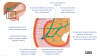
Slagter - Drawing Veins draining to the portal vein of the liver - English-Latin labels |
 |
opgobee |
Creative Commons Attribution-NonCommercial-ShareAlike |
2.473 |
8 |
|
Data item |
Radiopaedia - Drawing Development of the aortic arch and branches after 8 weeks - English labels |
 |
rva |
Creative Commons Attribution-NonCommercial-NoDerivs |
2.463 |
8 |
|
Data item |
Cenveo - Drawing Kidney and hilum in coronal section - English labels |
 |
rva |
Creative Commons Attribution |
2.462 |
9 |
|
Data item |
Leiden - Drawing Nociceptive pathways in the pelvis |
 |
opgobee |
Creative Commons Attribution-NonCommercial-ShareAlike |
2.462 |
3 |
|
Data item |
OpenStax AnatPhys fig.11.17 - The Diaphragm - English labels |
 |
Jorn IJkhout |
Creative Commons Attribution |
2.460 |
9 |
|
Data item |
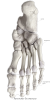
Anatomy Standard - Drawing Bones of foot: plantar view - Latin labels |
 |
rva |
Creative Commons Attribution-NonCommercial |
2.459 |
6 |
|
Anatomical Structure |
Linea obliqua mandibulae |
|
admin |
|
2.458 |
5 |
|
Data item |
Voorbeeld Opdracht vragen maken op AnatomyTOOL |
|
lumctest |
Creative Commons Attribution |
2.456 |
1 |
|
Data item |
3D Anatomy Lyon: The thumb (pollux) and opposition - video of 3D model |
|
rva |
Creative Commons Attribution-NonCommercial-NoDerivs |
2.453 |
10 |
|
Data item |
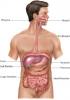
Servier - Drawing Overview digestive tract - no labels |
 |
rva |
Creative Commons Attribution |
2.452 |
5 |
|
Data item |
Radiopaedia - Drawing Cross-section of the penis - English labels |
 |
rva |
Creative Commons Attribution-NonCommercial-ShareAlike |
2.451 |
5 |
|
Data item |
KnowledgeWorks - Drawing Abdominal organs - English labels |
 |
rva |
Creative Commons Attribution-NonCommercial-ShareAlike |
2.447 |
4 |
|
Data item |
Sobotta 1909 fig.270 - superficial muscles of the arm, lateral view - English Labels |
 |
Student128 |
Public Domain |
2.443 |
3 |
|
Data item |
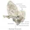
Anatomy Standard - Drawing Sphenoid bone: superior view - no labels |
 |
rva |
Creative Commons Attribution-NonCommercial |
2.442 |
16 |
|
Data item |
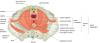
OpenStax AnatPhys fig.13.14 - Spinal Cord Cross Section - English labels |
 |
Jorn IJkhout |
Creative Commons Attribution |
2.440 |
9 |
|
Data item |
Transversus abdominis muscle |
|
opgobee |
Creative Commons Attribution-NonCommercial-ShareAlike |
2.438 |
3 |
|
Data item |
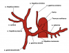
Universiteit Gent - Students - Drawing The celiac trunk (1) - Latin labels |
 |
sehellin |
Creative Commons Attribution-NonCommercial-ShareAlike |
2.433 |
11 |
|
Anatomical Structure |
Facies costalis scapulae |
|
admin |
|
2.428 |
3 |
|
Data item |
Anatomy Standard - Drawing Temporal bone: anterior view - Latin labels |
 |
rva |
Creative Commons Attribution-NonCommercial |
2.422 |
4 |
|
Data item |
3D Anatomy Lyon: Anatomy of the serratus anterior - video of 3D model |
|
rva |
Creative Commons Attribution-NonCommercial-NoDerivs |
2.415 |
5 |
|
Learning Path |
Quiz Maag - Kenmerken en Onderdelen (basis) |
|
tjscherphof |
Creative Commons Attribution |
2.414 |
0 |
|
Data item |
Anatomy Standard - Drawing Scapula: costal surface (anterior view) - no labels |
 |
rva |
Creative Commons Attribution-NonCommercial |
2.409 |
7 |
|
Data item |
OLI - Drawing The heart during systole and diastole - English labels |
 |
rva |
Creative Commons Attribution-NonCommercial-ShareAlike |
2.406 |
9 |
|
Data item |
Glandulae salivariae majores |
 |
koolstra |
Creative Commons Attribution-NonCommercial-ShareAlike |
2.395 |
2 |
|
Anatomical Structure |
Crista temporalis mandibulae |
|
admin |
|
2.394 |
2 |
|
Data item |
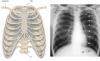
Slagter - Drawing and Chest X-ray of the ribs and clavicula - Latin labels |
 |
rva |
Creative Commons Attribution-NonCommercial-ShareAlike |
2.391 |
10 |
|
Learning Path |
Quiz Darm - Vascularisatie (basis) |
|
tjscherphof |
Creative Commons Attribution-NonCommercial-ShareAlike |
2.389 |
0 |
|
Data item |
OpenStax AnatPhys fig.11.17 - The Diaphragm and crura - English labels |
 |
admin |
Creative Commons Attribution |
2.386 |
2 |
|
Data item |
Sobotta 1909 fig.40 - The skull, lateral view - No labels |
 |
Student128 |
Public Domain |
2.381 |
1 |
|
Data item |
Radiopaedia - Drawing Brachial plexus - English labels |
 |
rva |
Creative Commons Attribution-NonCommercial-ShareAlike |
2.381 |
8 |
|
Data item |
JMarchn - Bile duct, pancreatic ducts, hepatopancreatic ampulla - Latin and English labels |
 |
opgobee |
Creative Commons Attribution-ShareAlike |
2.381 |
11 |
|
Data item |
Wikimedia Commons - anatomical resources |
|
opgobee |
This item is (on) an external site. The license as stated on that site holds. |
2.375 |
5 |
|
Data item |
Anatomy Standard - Drawing Hip bone (os coxae): medial view - no labels |
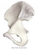 |
rva |
Creative Commons Attribution-NonCommercial |
2.374 |
6 |
|
Data item |
Slagter - Drawing Human oogenesis diagram with explanation - English labels |
 |
rva |
Creative Commons Attribution-NonCommercial-ShareAlike |
2.372 |
7 |
|
Data item |
OLI - Drawing Heart from anterior: flow directions - English labels |
 |
rva |
Creative Commons Attribution-NonCommercial-ShareAlike |
2.371 |
4 |
|
Data item |
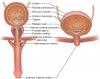
Cenveo - Drawing Male and female urinary tract - English labels |
 |
rva |
Creative Commons Attribution |
2.371 |
2 |
|
Data item |
Radiopaedia - Drawing Gyri and sulci: superior surface of brain - no labels |
 |
rva |
Creative Commons Attribution-NonCommercial-ShareAlike |
2.370 |
7 |
|
Data item |
Thunthu - 3D model Trachea and bronchi |
|
rva |
Creative Commons Attribution |
2.369 |
6 |
|
Data item |
Anatomy Standard - Drawing Mandibula: posterior view - Latin labels |
 |
rva |
Creative Commons Attribution-NonCommercial |
2.366 |
6 |
|
Data item |
Anatomy Standard - Drawing Atlas: superior aspect - Latin labels |
 |
rva |
Creative Commons Attribution-NonCommercial |
2.362 |
4 |
|
Data item |
ANATOMY GYM |
|
cjhaven |
Public Domain |
2.361 |
3 |
|
Data item |
Left inferior view of levator ani and external anal sphincter muscles -English labels |
 |
opgobee |
Creative Commons Attribution-NonCommercial-ShareAlike |
2.346 |
3 |
|
Data item |
Greensboro - 3D model Right os coxa |
|
rva |
Creative Commons Attribution |
2.345 |
5 |
|
Data item |
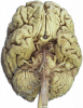
U.Br.Columbia - Photo Inferior brain: cranial nerves (dissection) - no labels |
 |
rva |
Creative Commons Attribution-NonCommercial-ShareAlike |
2.338 |
5 |
|
Data item |
Radiopaedia - Drawing Styloid apparatus - English labels |
 |
rva |
Creative Commons Attribution-NonCommercial-ShareAlike |
2.337 |
4 |
|
Data item |
MaastrichtUniversity - Student - Booklet with Anatomical Sketches Abdomen, Pelvis, Perineum and Genitals - Dutch labels |
|
Divine |
Creative Commons Attribution-NonCommercial-ShareAlike |
2.333 |
3 |
|
Data item |
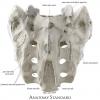
Anatomy Standard - Drawing Sacrum: posterior aspect - Latin labels |
 |
rva |
Creative Commons Attribution-NonCommercial |
2.333 |
7 |
|
Data item |
U.Br.Columbia - Drawing Innervation of submandibular and sublingual salivary glands - English labels |
 |
rva |
Creative Commons Attribution-NonCommercial-ShareAlike |
2.327 |
2 |
|
Data item |
OpenStax AnatPhys fig.14.15 - Structure of the Eye - English labels |
 |
Jorn IJkhout |
Creative Commons Attribution |
2.326 |
7 |
|
Data item |
Anatomy Standard - Drawing Superior aspect of cervical vertebra (C4) - Latin labels |
 |
rva |
Creative Commons Attribution-NonCommercial |
2.325 |
4 |
|
Data item |
3D Anatomy Lyon: The acromioclavicular joint - video of 3D model |
|
rva |
Creative Commons Attribution-NonCommercial-NoDerivs |
2.323 |
3 |
|
Data item |
Radiopaedia - CT-scan Hepatic segments: coronal section - labels |
 |
rva |
Creative Commons Attribution-NonCommercial-ShareAlike |
2.315 |
9 |
|
Data item |
Slagter - Drawing Lymph node levels of the neck - labels |
|
rva |
Creative Commons Attribution-NonCommercial-ShareAlike |
2.307 |
5 |
|
Data item |
U.Br.Columbia - Drawing Cross section through middle of thigh - English labels |
 |
rva |
Creative Commons Attribution-NonCommercial-ShareAlike |
2.304 |
1 |
|
Learning Path |
Quiz Galwegen (basis) |
|
tjscherphof |
Creative Commons Attribution-NonCommercial-ShareAlike |
2.301 |
0 |
|
Anatomical Structure |
Pars spongiosa |
|
admin |
|
2.295 |
1 |
|
Data item |
Radiopaedia - Drawing Main branches of the mandibular nerve - English labels |
 |
rva |
Creative Commons Attribution-NonCommercial-ShareAlike |
2.292 |
7 |