nid: 60216
Additional formats:
None available
Description:
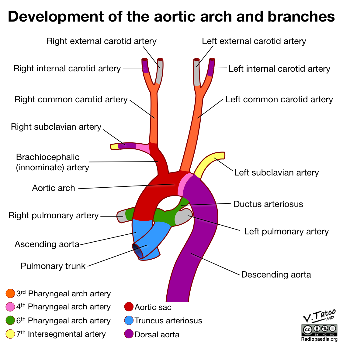
Development of the aortic arch and branches after 8 weeks. This diagram shows the development of a normal aortic arch from the embryonic pharyngeal arch arteries. English labels.
Case courtesy of Dr Vincent Tatco, Radiopaedia.org. From the case rID: 52192
Case courtesy of Dr Vincent Tatco, Radiopaedia.org. From the case rID: 52192
Anatomical structures in item:
Uploaded by: rva
Netherlands, Leiden – Leiden University Medical Center, Leiden University
Truncus pulmonalis
Aorta ascendens
Arteriae pulmonalis dextra
Arcus aortae
Truncus brachiocephalicus
Arteria subclavia dextra
Arteria carotis communis
Arteria carotis interna
Arteria carotis externa
Arteria subclavia
Ligamentum arteriosum (Ductus arteriosus)
Aorta
Arteria pulmonalis sinistra
Aorta descendens
Creator(s)/credit: Dr Vincent Tatco
Requirements for usage
You are free to use this item if you follow the requirements of the license:  View license
 View license
 If you use this item you should credit it as follows:
- For usage in print - copy and paste the line below:
- For digital usage (e.g. in PowerPoint, Impress, Word, Writer) - copy and paste the line below (optionally add the license icon):
"Radiopaedia - Drawing Development of the aortic arch and branches after 8 weeks - English labels" at AnatomyTOOL.org by Vincent Tatco, license: Creative Commons Attribution-NonCommercial-NoDerivs


Comments