nid: 62596
Additional formats:
None available
Description:
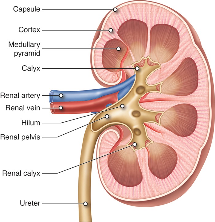
Kidney and hilum in coronal section. This image shows a cross section of the left kidney. English labels.
Retrieved from Anatomy & Physiology by Open Learning Initiative (CC BY-NC-SA).
Retrieved from Anatomy & Physiology by Open Learning Initiative (CC BY-NC-SA).
Anatomical structures in item:
Uploaded by: rva
Netherlands, Leiden – Leiden University Medical Center, Leiden University
Ren (Nephros)
Capsula fibrosa renis
Cortex renalis
Pyramides renales
Pyramis medullae oblongatae
Superior major calyx
Intermediate major calyx
Inferior major calyx
Calices renales majores
Arteria renalis
Venae renales
Hilum renale
Pelvis renalis
Ureter
Creator(s)/credit: Cenveo
Requirements for usage
You are free to use this item if you follow the requirements of the license:  View license
 View license
 If you use this item you should credit it as follows:
- For usage in print - copy and paste the line below:
- For digital usage (e.g. in PowerPoint, Impress, Word, Writer) - copy and paste the line below (optionally add the license icon):
"Cenveo - Drawing Kidney and hilum in coronal section - English labels" at AnatomyTOOL.org by Cenveo, license: Creative Commons Attribution
"Cenveo - Drawing Kidney and hilum in coronal section - English labels" by Cenveo, license: CC BY


Comments