|
Data item |
Maastricht - 3D models development of aortic arch arteries |
|
chulsman |
Creative Commons Attribution-NonCommercial-ShareAlike |
1.224 |
5 |
|
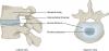
Data item |
Saggital section male pelvis with filled bladder |
 |
lumcanatomy |
Creative Commons Attribution-NonCommercial-ShareAlike |
1.224 |
2 |
|
Data item |
Cenveo - Drawing Structures of the Vestibular Apparatus - English labels |
 |
rva |
Creative Commons Attribution |
1.224 |
6 |
|
Data item |
Dundee - Drawing Abduction of the hip: muscles and tendons seen from posterior - English labels |
 |
rva |
Creative Commons Attribution-NonCommercial-NoDerivs |
1.222 |
2 |
|
Data item |
OLI - Table Comparison of various blood vessels - English labels |
 |
rva |
Creative Commons Attribution-NonCommercial-ShareAlike |
1.222 |
2 |
|
Learning Path |
Quiz Perineum - Kenmerken en onderdelen (gevorderd) |
|
opgobee |
Creative Commons Attribution-NonCommercial-ShareAlike |
1.221 |
0 |
|
Data item |
Cavitas oris |
 |
koolstra |
Creative Commons Attribution-NonCommercial-ShareAlike |
1.221 |
4 |
|
Data item |
OpenStax AnatPhys fig.21.4 - Lymphatic Trunks and Ducts System - English labels |
 |
Jorn IJkhout |
Creative Commons Attribution |
1.220 |
2 |
|
Data item |
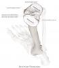
Anatomy Standard - Drawing Right ulna: posterior view - Latin labels |
 |
rva |
Creative Commons Attribution-NonCommercial |
1.220 |
2 |
|
Learning Path |
Quiz Anatomie van Pijn A - Structuren en Systemen 2 |
|
opgobee |
Creative Commons Attribution-NonCommercial-ShareAlike |
1.220 |
1 |
|
Data item |
Anatomy Standard - Drawing Parietal bone: external (lateral) view - Latin labels |
 |
rva |
Creative Commons Attribution-NonCommercial |
1.220 |
4 |
|
Data item |
KnowledgeWorks - Drawing Abdominal organs and detail of anal canal - English labels |
 |
rva |
Creative Commons Attribution |
1.218 |
3 |
|
Data item |
Anatomy Standard - Drawing Temporal bone: lateral view - no labels |
 |
rva |
Creative Commons Attribution-NonCommercial |
1.218 |
7 |
|
Data item |
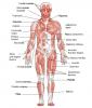
Human Biology fig. 1.19 - Global image of some important skeletal muscles - English labels |
 |
rva |
Creative Commons Attribution-NonCommercial |
1.218 |
1 |
|
Data item |
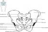
OpenStax AnatPhys fig.8.12 - Pelvis - English labels |
 |
Jorn IJkhout |
Creative Commons Attribution |
1.218 |
7 |
|
Data item |
Lingua, plana frontalia |
 |
koolstra |
Creative Commons Attribution-NonCommercial-ShareAlike |
1.217 |
4 |
|
Data item |
Dundee - Drawing Cartilage Anatomy of the Larynx - English labels |
 |
rva |
Creative Commons Attribution-ShareAlike |
1.216 |
3 |
|
Anatomical Structure |
Facies posterior scapulae |
|
admin |
|
1.215 |
0 |
|
Data item |
OpenStax AnatPhys fig. 11.23 - Muscles that Move the Humerus - no labels |
 |
rva |
Creative Commons Attribution |
1.214 |
5 |
|
Learning Path |
Quiz Aorta Abdominalis Takken (gevorderd) |
|
tjscherphof |
Creative Commons Attribution |
1.213 |
2 |
|
Data item |
arteria vertebralis |
 |
aherrler |
Creative Commons Attribution-NonCommercial-ShareAlike |
1.212 |
4 |
|
Learning Path |
Quiz Nier - Vascularisatie (gevorderd) |
|
tjscherphof |
Creative Commons Attribution-NonCommercial-ShareAlike |
1.212 |
1 |
|
Data item |
thorax lateral |
 |
aherrler |
Creative Commons Attribution-ShareAlike |
1.212 |
7 |
|
Data item |
Adominal wall -introduction |
|
opgobee |
Creative Commons Attribution-NonCommercial-ShareAlike |
1.210 |
3 |
|
Anatomical Structure |
Extremitas sternalis claviculae |
|
admin |
|
1.209 |
0 |
|
Anatomical Structure |
Radix linguae |
|
admin |
|
1.208 |
1 |
|
Data item |
OpenStax AnatPhys fig.1.13 - Directional Terms Applied to the Human Body - English labels |
 |
opgobee |
Creative Commons Attribution |
1.207 |
3 |
|
Anatomical Structure |
Carunculae hymenales |
|
admin |
|
1.207 |
1 |
|
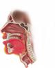
Data item |
Cenveo - Drawing Lateral view of the nose and mid-sagittal section through the nasal cavity - English labels |
 |
rva |
Creative Commons Attribution |
1.203 |
4 |
|
Anatomical Structure |
Ligamentum ovarii proprium |
|
admin |
|
1.203 |
3 |
|
Data item |
3D Anatomy Lyon: Vibration of the vocal folds - video of 3D model |
|
rva |
Creative Commons Attribution-NonCommercial-NoDerivs |
1.202 |
5 |
|
Data item |
OpenStax AnatPhys fig.11.32 - Muscles of the Leg that Move the Foot and Toes - English labels |
 |
Jorn IJkhout |
Creative Commons Attribution |
1.201 |
2 |
|
Data item |
NYSORA - Drawing Innervation of forearm - English labels |
 |
rva |
Creative Commons Attribution-NonCommercial-NoDerivs |
1.198 |
1 |
|
Data item |
Froodrice - 3D model Whole Sphenoid bone |
|
rva |
Creative Commons Attribution-ShareAlike |
1.198 |
2 |
|
Data item |
Goodwin - Drawing Cross section of spinal cord - English labels |
 |
rva |
Creative Commons Attribution-NonCommercial-ShareAlike |
1.195 |
6 |
|
Data item |
Anatomy Standard - Drawing Superior articular surface of tibia - Latin labels |
 |
rva |
Creative Commons Attribution-NonCommercial |
1.194 |
4 |
|
Data item |
CT abdomen transvers 1 |
 |
aherrler |
Creative Commons Attribution-NonCommercial-ShareAlike |
1.194 |
5 |
|
Data item |
Leiden - Student - Drawing Anatomy of the heart - Numbered labels and Latin answers |
 |
Flynn Post |
Creative Commons Attribution-ShareAlike |
1.192 |
5 |
|
Data item |
Radiopaedia - Drawing Fat pads and bursae of the knee - English labels |
 |
rva |
Creative Commons Attribution-NonCommercial-ShareAlike |
1.191 |
2 |
|
Data item |
Sobotta 1906 fig.435 - Larynx, frontal section - English labels |
 |
Student128 |
Public Domain |
1.191 |
3 |
|
Data item |
LadyofHats - Drawing Bones, sutures and foramens of the skull - English labels |
 |
rva |
Public Domain |
1.189 |
6 |
|
Data item |
Anatomy Standard - Drawing Hyoid bone: anterior view - no labels |
 |
rva |
Creative Commons Attribution-NonCommercial |
1.189 |
5 |
|
Data item |
BlueLink - 3D model First right rib |
|
rva |
Creative Commons Attribution-NonCommercial-NoDerivs |
1.188 |
2 |
|
Data item |
3D Anatomy Lyon: The ligaments of the spinal column - video of 3D model |
|
rva |
Creative Commons Attribution-NonCommercial-NoDerivs |
1.188 |
2 |
|
Data item |
Elon - 3D model Occipital Bone |
|
rva |
Creative Commons Attribution |
1.187 |
6 |
|
Data item |
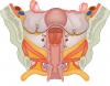
Coronal section of the female pelvis showing the cervix, vagina, cardinal ligament and levator ani muscle – no labels |
 |
Siem Zethof |
Creative Commons Attribution-NonCommercial-ShareAlike |
1.187 |
5 |
|
Data item |
OpenStax AnatPhys fig.11.17 - The Diaphragm - no labels |
 |
opgobee |
Creative Commons Attribution |
1.187 |
4 |
|
Data item |
U.Br.Columbia - Drawing Sagittal view of the mediastinum - English labels |
 |
rva |
Creative Commons Attribution-NonCommercial-ShareAlike |
1.185 |
3 |
|
Data item |
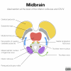
Radiopaedia - Drawing Midbrain at level of inferior colliculus and trochlear nerve - English labels |
 |
rva |
Creative Commons Attribution-NonCommercial-ShareAlike |
1.184 |
2 |
|
Anatomical Structure |
Vena brachiocephalica |
|
admin |
|
1.184 |
1 |
|
Anatomical Structure |
Duodenum |
|
admin |
|
1.184 |
1 |
|
Data item |
MedicalGraphics - Drawing Pancreas and duodenum - no labels |
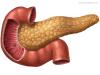 |
rva |
Creative Commons Attribution-NoDerivatives |
1.183 |
2 |
|
Data item |
Slagter - Drawing Biliary system - Latin and English labels |
 |
opgobee |
Creative Commons Attribution-NonCommercial-ShareAlike |
1.182 |
1 |
|
Data item |
U.Br.Columbia - Drawing First layer of muscles of the sole - English labels |
 |
rva |
Creative Commons Attribution-NonCommercial-ShareAlike |
1.182 |
2 |
|
Data item |
Radiopaedia - Drawing Origins of Arnold's and Jacobson's nerve - English labels |
 |
rva |
Creative Commons Attribution-NonCommercial-ShareAlike |
1.182 |
4 |
|
Data item |
Sobotta 1909 fig.208 - male pelvis with ligaments, anterior view - English Labels |
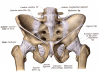 |
Student128 |
Public Domain |
1.181 |
5 |
|
Data item |
OpenStax AnatPhys fig.20.37 - Head and Neck Veins - English labels |
 |
Jorn IJkhout |
Creative Commons Attribution |
1.180 |
6 |
|
Data item |
OpenStax AnatPhys fig.13.9 - Frontal Section Basal Nuclei - English labels |
 |
Jorn IJkhout |
Creative Commons Attribution |
1.180 |
7 |
|
Learning Path |
Quiz Nier - Rondom de Nier (basis) |
|
tjscherphof |
Creative Commons Attribution-NonCommercial-ShareAlike |
1.180 |
0 |
|
Data item |
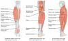
Sobotta 1909 fig.306 - anterior muscles of the leg - English Labels |
 |
Student128 |
Public Domain |
1.178 |
1 |
|
Anatomical Structure |
Foramina papillaria renalis |
|
admin |
|
1.177 |
2 |
|
Learning Path |
Quiz Anatomie van Pijn A - Structuren en Systemen 3 |
|
opgobee |
Creative Commons Attribution-NonCommercial-ShareAlike |
1.177 |
2 |
|
Data item |
Lynch - Drawing Anterior view of cranium and mandible - no labels |
 |
rva |
Creative Commons Attribution |
1.175 |
2 |
|
Anatomical Structure |
Collum chirurgicum humeri |
|
admin |
|
1.174 |
5 |
|
Data item |
U.Br.Columbia - Drawing Corpus callosum from lateral - English labels |
 |
rva |
Creative Commons Attribution-NonCommercial-ShareAlike |
1.173 |
6 |
|
Data item |
Sobotta 1909 fig.283 - muscles and ligaments of the palm - no labels |
 |
lumcanatomy |
Creative Commons Attribution-ShareAlike |
1.173 |
4 |
|
Anatomical Structure |
Facies intervertebralis |
|
admin |
|
1.172 |
0 |
|
Learning Path |
Quiz Thoraxwand - Bloedvaten en zenuwen |
|
opgobee |
Creative Commons Attribution-NonCommercial-ShareAlike |
1.171 |
0 |
|
Anatomical Structure |
Ostium urethrae internum (Urethra feminina) |
|
admin |
|
1.170 |
4 |
|
Anatomical Structure |
Crista colli costae |
|
admin |
|
1.169 |
0 |
|
Data item |
Leiden - Student - Drawing Coronary vessels - Numbered labels |
 |
Flynn Post |
Creative Commons Attribution-ShareAlike |
1.168 |
3 |
|
Data item |
Sobotta 1909 fig.628 - Fissures and convulsions of the cerebral cortex, superior view - coloured, no labels |
 |
rva |
Creative Commons Attribution-ShareAlike |
1.168 |
4 |
|
Data item |
Servier - Drawing Superficial muscles anterior view - no labels |
 |
rva |
Creative Commons Attribution |
1.168 |
12 |
|
Data item |
Radiopaedia - Drawing Superficial venous sinuses and cerebral veins - English labels |
 |
rva |
Creative Commons Attribution-NonCommercial-ShareAlike |
1.167 |
1 |
|
Data item |
BlueLink - 3D model Fifth Lumbar Vertebra (L5) |
|
rva |
Creative Commons Attribution-NonCommercial-NoDerivs |
1.167 |
4 |
|
Data item |
RCSI - Drawing Thyroid and parathyroid glands and vasculature - English labels |
 |
rva |
Creative Commons Attribution-NonCommercial-ShareAlike |
1.166 |
4 |
|
Anatomical Structure |
Truncus thyrocervicalis |
|
admin |
|
1.166 |
0 |
|
Data item |
human brain, 3D |
|
aherrler |
Creative Commons Attribution-NonCommercial-ShareAlike |
1.165 |
1 |
|
Learning Path |
Heart and Vessels Entrance exam #1 |
|
leo.koehler |
Creative Commons Attribution-NonCommercial-ShareAlike |
1.164 |
1 |
|
Data item |
Thunthu - 3D model Bones of the lower limb |
|
rva |
Creative Commons Attribution |
1.164 |
8 |
|
Anatomical Structure |
Musculi linguae |
|
admin |
|
1.163 |
2 |
|
Learning Path |
GI-Tract Entrance exam #2 |
|
leo.koehler |
Creative Commons Attribution-NonCommercial-ShareAlike |
1.162 |
0 |
|
Learning Path |
Quiz LUMC Endoscopieverpleegkundigen vascularisatie spijsverteringsstelsel |
|
opgobee |
Creative Commons Attribution-NonCommercial-ShareAlike |
1.161 |
0 |
|
Data item |
Lynch - Drawing Muscles of the head - no labels |
 |
rva |
Creative Commons Attribution |
1.161 |
7 |
|
Data item |
Slagter - Drawing Muscles of arm: anterior view - no labels |
 |
rva |
Creative Commons Attribution-NonCommercial-ShareAlike |
1.160 |
2 |
|
Data item |
Servier - Drawing Layers of the heart wall - English labels |
 |
rva |
Creative Commons Attribution |
1.160 |
4 |
|
Data item |
Leiden, Maas Photo 30 - Female pelvis view from anterior (plastination specimen) - number labels |
 |
rva |
Creative Commons Attribution-NonCommercial-ShareAlike |
1.160 |
4 |
|
Data item |
Anatomy Standard - Drawing Sphenoid bone: anterior view - no labels |
 |
rva |
Creative Commons Attribution-NonCommercial |
1.160 |
11 |
|
Data item |
OLI - Drawing Detail of muscle fiber - English labels |
 |
rva |
Creative Commons Attribution-NonCommercial-ShareAlike |
1.159 |
7 |
|
Anatomical Structure |
Facies temporalis ossis frontalis |
|
admin |
|
1.159 |
3 |
|
Data item |
Sobotta 1909 fig.726 - Sympathetic trunk and vagus nerve, thoracic (and abdominal) portion - no labels |
 |
rva |
Creative Commons Attribution-ShareAlike |
1.158 |
2 |
|
Anatomical Structure |
Caput costae |
|
admin |
|
1.157 |
1 |
|
Data item |
Slagter - Drawing Head and neck: sagittal section - no labels |
 |
rva |
Creative Commons Attribution-NonCommercial-ShareAlike |
1.157 |
3 |
|
Data item |
RCSI - Drawing Breast cross-section - English labels |
 |
rva |
Creative Commons Attribution-NonCommercial-ShareAlike |
1.157 |
6 |
|
Data item |
OpenStax AnatPhys fig.7.24 - Intervertebral Disk - English labels |
 |
Jorn IJkhout |
Creative Commons Attribution |
1.156 |
2 |
|
Data item |
OpenStax AnatPhys fig.25.13(b) - Juxtaglomerular Apparatus and Glomerulus - English labels |
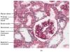 |
Jorn IJkhout |
Creative Commons Attribution |
1.155 |
4 |
|
Anatomical Structure |
Musculi capitis |
|
admin |
|
1.155 |
0 |
|
Learning Path |
Quiz Perineum - Kenmerken en onderdelen (basis) |
|
opgobee |
Creative Commons Attribution-NonCommercial-ShareAlike |
1.155 |
0 |
|
Data item |
Leiden - Photo Duodenum and pancreas dissection specimen - no labels |
 |
opgobee |
Creative Commons Attribution-NonCommercial-ShareAlike |
1.153 |
5 |
|
Anatomical Structure |
Linea axillaris anterior |
|
admin |
|
1.153 |
2 |