
nid: 59742
Additional formats:
None available
Description:
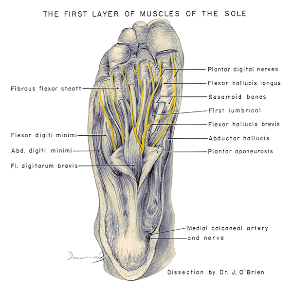
First layer of muscles of the sole. The first layer of muscles of the sole of the foot can be seen. The innervating nerves are also visible. English labels.
Retrieved from website Clinical Anatomy of the University of British Columbia.
Retrieved from website Clinical Anatomy of the University of British Columbia.
Anatomical structures in item:
Uploaded by: rva
Netherlands, Leiden – Leiden University Medical Center, Leiden University
Pes
Planta
Flexor digiti minimi brevis of foot
Musculus flexor digitorum brevis
Aponeurosis plantaris
Musculus abductor hallucis
Musculus flexor hallucis brevis
Os sesamoideum
Musculus flexor hallucis longus
Creator(s)/credit: Department of Anatomy, University of British Columbia, UBC; Dr. J.P O'Brien, UBC
Requirements for usage
You are free to use this item if you follow the requirements of the license:  View license
View license If you use this item you should credit it as follows:
- For usage in print - copy and paste the line below:
- For digital usage (e.g. in PowerPoint, Impress, Word, Writer) - copy and paste the line below (optionally add the license icon):
"U.Br.Columbia - Drawing First layer of muscles of the sole - English labels" at AnatomyTOOL.org by Department of Anatomy, University of British Columbia, UBC and J.P O'Brien, UBC, license: Creative Commons Attribution-NonCommercial-ShareAlike. Created for: Department of Anatomy (now Department of Cellular and Physiological Sciences) at the University of British Columbia. Source: website Clinical Anatomy, http://www.clinicalanatomy.ca
"U.Br.Columbia - Drawing First layer of muscles of the sole - English labels" by Department of Anatomy, University of British Columbia, UBC and J.P O'Brien, UBC, license: CC BY-NC-SA. Created for: Department of Anatomy (now Department of Cellular and Physiological Sciences) at the University of British Columbia. Source: website Clinical Anatomy, http://www.clinicalanatomy.ca


Comments