
nid: 63543
Additional formats:
- Presentation slide Leiden - Drawing Cross section upper abdomen with peritoneal lining and Mattox manoevre - labels.pptx, *.pptx, 347kB, Powerpoint version with labels, for editing
Description:
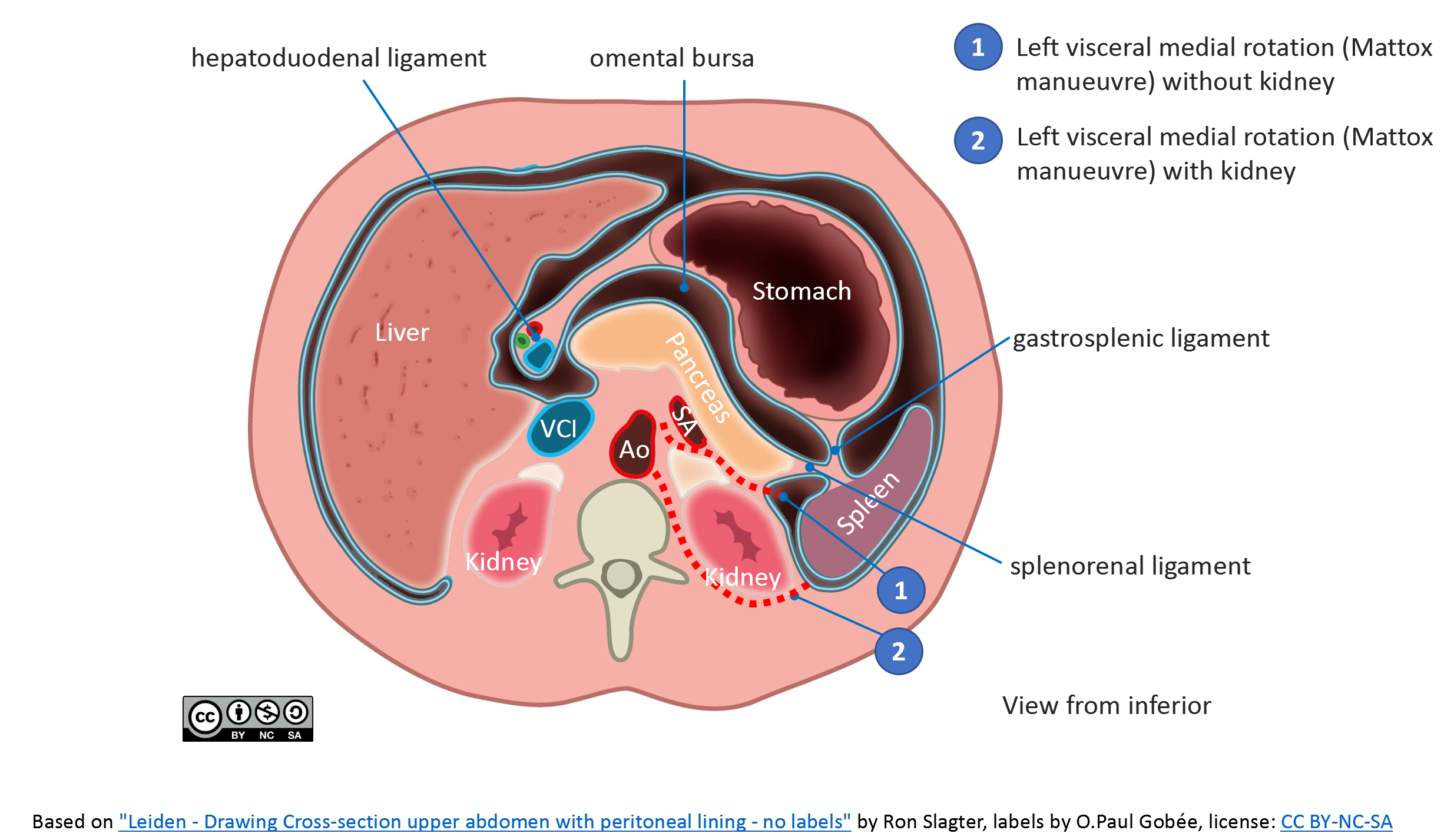
Transverse section of the abdomen (caudal view) with peritoneal lining in blue. This drawing was made for the MOOC "Anatomy of the Abdomen and Pelvis; a journey from basis to clinic" English labels.
The dissection routes of the visceral medial rotation (Mattox manoevre) with and without mobilizing the kidney, are indicated by red dashed lines.
VCI: Vena Cava inferior, Ao: aorta; SA: Splenic artery
This drawing was made for the MOOC "Anatomy of the Abdomen and Pelvis; a journey from basis to clinic" Dutch labels.
Anatomical structures in item:
Uploaded by: admin
Netherlands, Leiden – Leiden University Medical Center, Leiden University
Hepar
Pancreas
Splen
Ventriculus
Ren (Nephros)
Glandula suprarenalis
Ligamentum hepatoduodenale
Aorta abdominalis
Vena cava inferior
Peritoneum
Ligamentum gastrolienale
Ligamentum splenorenale
Bursa omentalis
Creator(s)/credit: Ron Slagter NZIMBI, medical illustrator; O. Paul Gobée MD, anatomists, labels, LUMC
Requirements for usage
You are free to use this item if you follow the requirements of the license:  View license
View license If you use this item you should credit it as follows:
- For usage in print - copy and paste the line below:
- For digital usage (e.g. in PowerPoint, Impress, Word, Writer) - copy and paste the line below (optionally add the license icon):
"Leiden - Drawing Cross section upper abdomen with peritoneal lining and Mattox manoevre - English labels" at AnatomyTOOL.org by Ron Slagter and O. Paul Gobée, LUMC, license: Creative Commons Attribution-NonCommercial-ShareAlike
"Leiden - Drawing Cross section upper abdomen with peritoneal lining and Mattox manoevre - English labels" by Ron Slagter and O. Paul Gobée, LUMC, license: CC BY-NC-SA


Comments