
nid: 62324
Additional formats:
None available
Description:
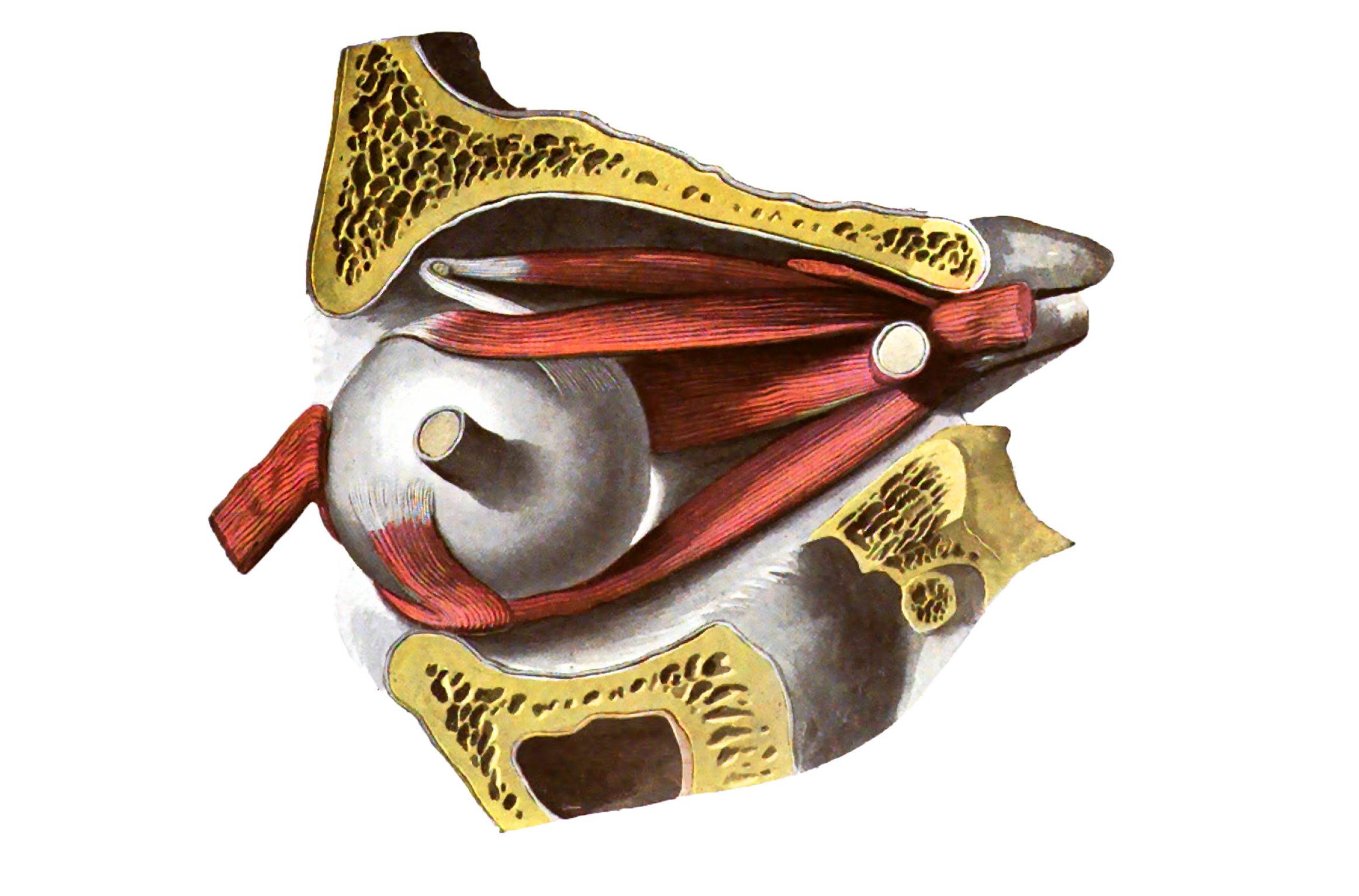
Ocular muscles, lateral view: preparation of fig.748. The optic nerve and the rectus lateralis have been divided and the eyeball has been rotated so that the stump of the optic nerve is directed laterally. Greater portion of the superior levator palpebrae has been removed.
From 'Atlas and Textbook of Human Anatomy', 1911 (?), Vol. 3, fig.749, by Johannes Sobotta and J. Playfair McMurrich. Artist: K. Hajek. Retrieved from Sobotta's Anatomy plates at Wikimedia.
Image editing by dream_studio3.
From 'Atlas and Textbook of Human Anatomy', 1911 (?), Vol. 3, fig.749, by Johannes Sobotta and J. Playfair McMurrich. Artist: K. Hajek. Retrieved from Sobotta's Anatomy plates at Wikimedia.
Image editing by dream_studio3.
Anatomical structures in item:
Uploaded by: rva
Netherlands, Leiden – Leiden University Medical Center, Leiden University
Bulbus oculi
Musculus rectus superior
Musculus rectus lateralis
Musculus rectus inferior
Musculus obliquus inferior
Musculus levator palpebrae superioris
Os frontale
Anulus tendineus communis
Nervus opticus
Os sphenoidale
Fossa infratemporalis
Fissura orbitalis inferior
Sinus maxillaris
Maxilla
Cornea
Musculus obliquus superior
Musculus rectus medialis
Margo supraorbitalis
Creator(s)/credit: Prof.dr. Johannes Sobotta, anatomist; dream_studio3 BA, image editing
Requirements for usage
You are free to use this item if you follow the requirements of the license:  View license
View license If you use this item you should credit it as follows:
- For usage in print - copy and paste the line below:
- For digital usage (e.g. in PowerPoint, Impress, Word, Writer) - copy and paste the line below (optionally add the license icon):
"Sobotta 1911 fig.749 - Ocular muscles, lateral view - no labels" at AnatomyTOOL.org by Johannes Sobotta and dream_studio3, license: Creative Commons Attribution-ShareAlike


Comments