
nid: 58306
Additional formats:
None available
Description:
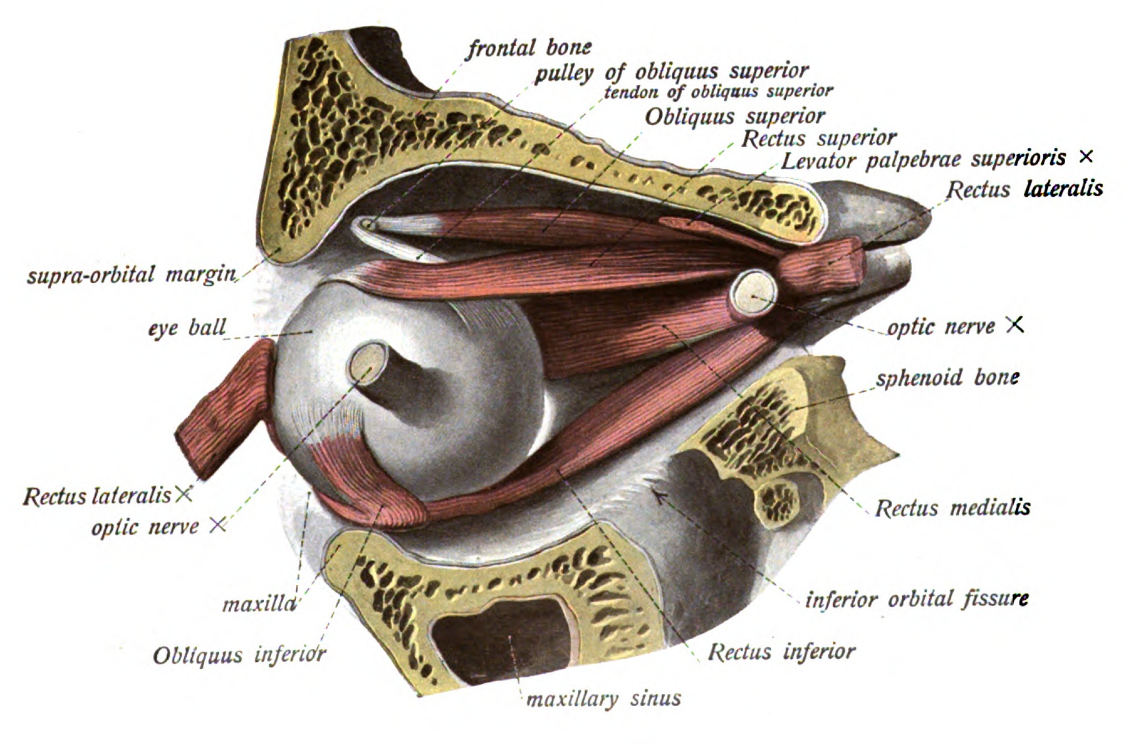
Ocular muscles, lateral view: preparation of fig.748. The optic nerve and the rectus lateralis have been divided and the eyeball has been rotated so that the stump of the optic nerve is directed laterally. Greater portion of the superior levator palpebrae has been removed. English labels.
From 'Atlas and Textbook of Human Anatomy', 1911 (?), Vol. 3, fig.749, by Johannes Sobotta and J. Playfair McMurrich. Artist: K. Hajek. Retrieved from Sobotta's Anatomy plates at Wikimedia.
From 'Atlas and Textbook of Human Anatomy', 1911 (?), Vol. 3, fig.749, by Johannes Sobotta and J. Playfair McMurrich. Artist: K. Hajek. Retrieved from Sobotta's Anatomy plates at Wikimedia.
Anatomical structures in item:
Uploaded by: Student128
Netherlands, Leiden – Leiden University Medical Center, Leiden University
Bulbus oculi
Musculus rectus superior
Musculus rectus lateralis
Musculus rectus inferior
Musculus obliquus inferior
Musculus levator palpebrae superioris
Os frontale
Anulus tendineus communis
Nervus opticus
Os sphenoidale
Fossa infratemporalis
Fissura orbitalis inferior
Sinus maxillaris
Maxilla
Cornea
Musculus obliquus superior
Musculus rectus medialis
Margo supraorbitalis
Creator(s)/credit: Prof.dr. Johannes Sobotta, anatomist
Requirements for usage
You are free to use this item.  Read more
Read more This item is in the Public Domain because its copyright has expired. You are not required to credit its creators when you use it. Nevertheless, it is adviced to do so. First, it is academically correct to pay tribute to the creators. Second, items of unknown origin might be classified as 'copyright infringement' by copyright controlling bodies, with possible resulting bills. Stating the item's source will prevent this. You can use the following text:
- For usage in print - copy and paste the line below:
- For digital usage (e.g. in PowerPoint, Impress, Word, Writer) - copy and paste the line below (optionally add the icon):
"Sobotta 1911 fig.749 - Ocular muscles, lateral view - English labels" at AnatomyTOOL.org by Johannes Sobotta is in the Public Domain.
"Sobotta 1911 fig.749 - Ocular muscles, lateral view - English labels" by Johannes Sobotta is in the Public Domain.


Comments