
nid: 62368
Additional formats:
None available
Description:
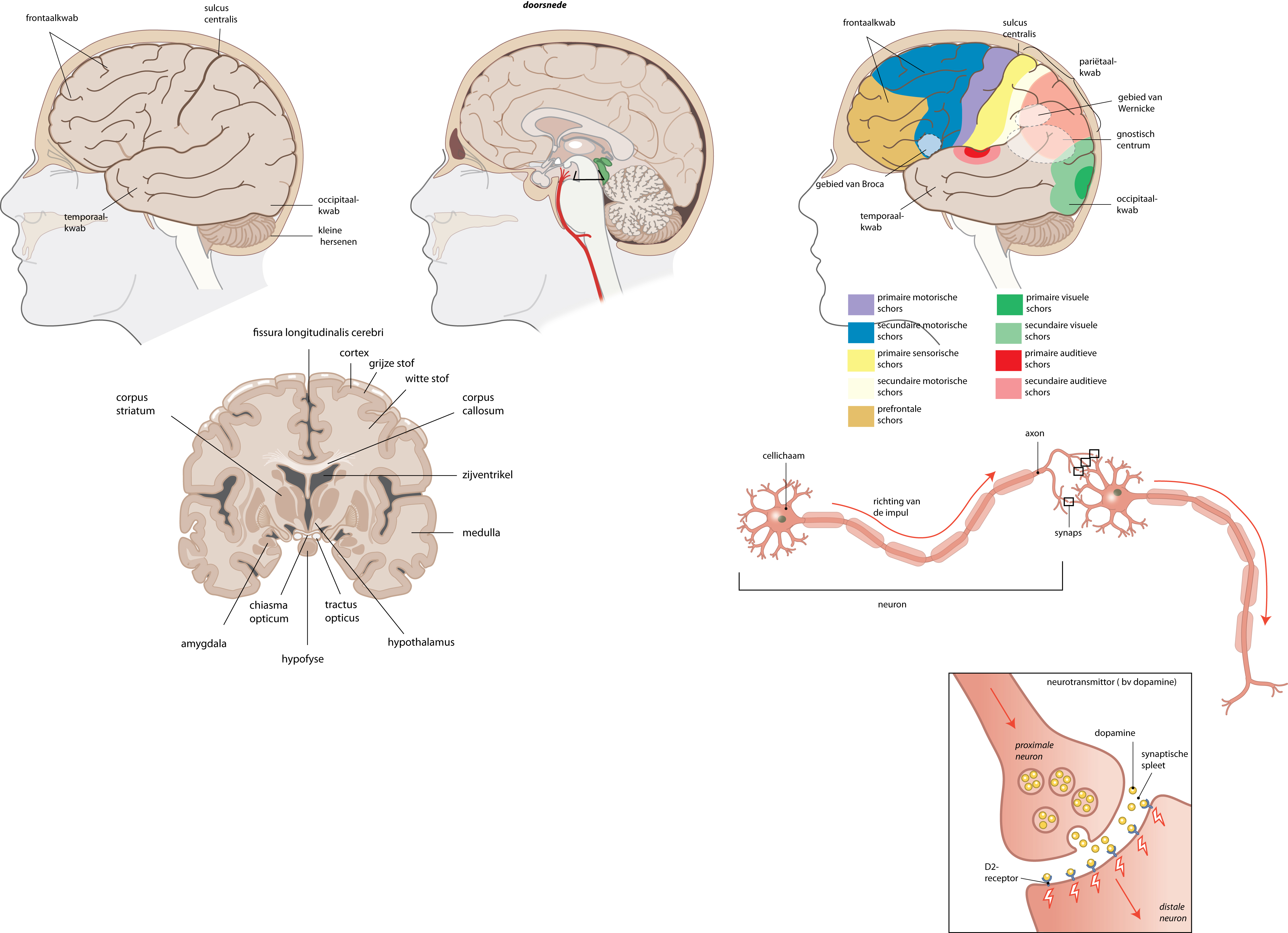
Anatomy of the brain and neurons. The three drawings on the left are a lateral view of the brain, a sagittal section of the brain, and a coronal section of the brain. The three drawings on the right are from upper to lower: location of different cortexes and areas of the brain, anatomy of a neuron, and a synapse. Dutch labels.
Anatomical structures in item:
Uploaded by: rva
Netherlands, Leiden – Leiden University Medical Center, Leiden University
Truncus encephali
Encephalon
Cerebellum
Corpus striatum
Corpus amygdaloideum
Chiasma opticum
Glandula pituitaria
Tractus opticus
Hypothalamus
Pars centralis ventriculi lateralis
Corpus callosum
Substantia alba medullae oblongatae
Substantia grisea
Cortex cerebelli
Fissura longitudinalis cerebri
Lobus frontalis
Gyrus frontalis inferior
Lobus temporalis
Sulcus centralis cerebri
Lobus parietalis
Lobus occipitalis
Lobus frontalis
Neuron
Synapsis
Creator(s)/credit: Ron Slagter NZIMBI, medical illustrator
Requirements for usage
You are free to use this item if you follow the requirements of the license:  View license
View license If you use this item you should credit it as follows:
- For usage in print - copy and paste the line below:
- For digital usage (e.g. in PowerPoint, Impress, Word, Writer) - copy and paste the line below (optionally add the license icon):
"Slagter - Drawing Anatomy of the brain and neurons - Dutch labels" at AnatomyTOOL.org by Ron Slagter, license: Creative Commons Attribution-NonCommercial-ShareAlike


Comments