
nid: 63531
Additional formats:
- Calot's triangle slide.pptx, *.pptx, 387kB, Powerpoint version for editing
Description:
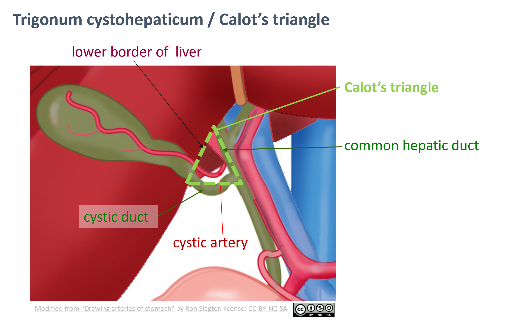
Calot's triangle is an aid in correct identification of the structures in the porta hepatis, in cholecystectomy.
Nowadays, it is considered to be the triangle formed by the common hepatic duct, the cystic duct and the lower border of the liver.
The cystic artery is then found within the triangle.
Originally, in Calot's description, the cystic artery was the third side of the triangle, instead of the lower border of the liver.
Anatomical structures in item:
Uploaded by: opgobee
Netherlands, Leiden – Leiden University Medical Center, Leiden University
Trigonum cystohepaticum
Ductus cysticus
Ductus hepaticus communis
Arteria cystica
Creator(s)/credit: Ron Slagter NZIMBI, medical illustrator; O. Paul Gobée MD, anatomist, LUMC
Requirements for usage
You are free to use this item if you follow the requirements of the license:  View license
View license If you use this item you should credit it as follows:
- For usage in print - copy and paste the line below:
- For digital usage (e.g. in PowerPoint, Impress, Word, Writer) - copy and paste the line below (optionally add the license icon):
"Leiden - Drawing Calot's triangle - English labels" at AnatomyTOOL.org by Ron Slagter and O. Paul Gobée, LUMC, license: Creative Commons Attribution-NonCommercial-ShareAlike
"Leiden - Drawing Calot's triangle - English labels" by Ron Slagter and O. Paul Gobée, LUMC, license: CC BY-NC-SA


Comments