
nid: 63808
Additional formats:
None available
Description:
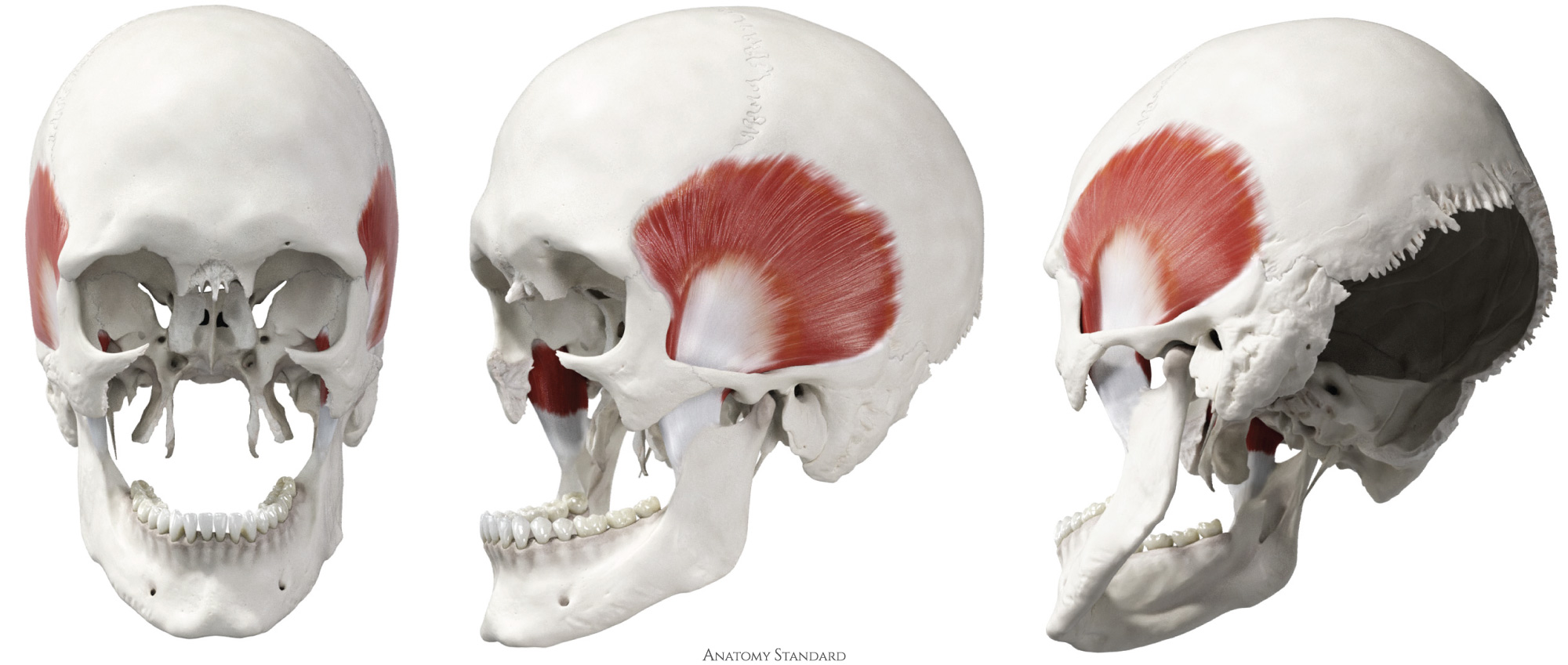
Temporal muscle: anterior, lateral and posterolateral view. Origin: temporal fossa. Insertion: coronoid process of the mandible, pterygomandibular raphe. Function: elevate (close) lower jaw. Dorsal fibers are involved in the retrusion or backward movement of the mandible. Version without labels.
Image and description retrieved from Anatomy Standard, page Masticatory Muscles.
Image and description retrieved from Anatomy Standard, page Masticatory Muscles.
Anatomical structures in item:
Uploaded by: rva
Netherlands, Leiden – Leiden University Medical Center, Leiden University
Musculus temporalis
Creator(s)/credit: Jānis Šavlovskis MD, PhD, Assistant Professor; Kristaps Raits, 3D generalist
Requirements for usage
You are free to use this item if you follow the requirements of the license:  View license
View license If you use this item you should credit it as follows:
- For usage in print - copy and paste the line below:
- For digital usage (e.g. in PowerPoint, Impress, Word, Writer) - copy and paste the line below (optionally add the license icon):
"Anatomy Standard - Temporal muscle: anterior, lateral and posterolateral view - no labels" at AnatomyTOOL.org by Jānis Šavlovskis and Kristaps Raits, license: Creative Commons Attribution-NonCommercial


Comments