

nid: 59520
Additional formats:
None available
Description:
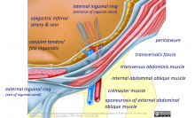
Label edit file of Abdominal wall layers of the inguinal canal, semi 3D exploded view. You can use this presentation file to easily create images with your own labels or use it as part of your own presentations. This file was used to create the images 'Abdominal wall layers of the inguinal canal, semi 3D exploded view - English labels'
The base image is 'Abdominal wall layers of the inguinal canal, semi 3D exploded view, no labels' illustration by Bas Blankevoort. Labels by Paul Gobée, MD. The image shows the abdominal wall layers forming the inguinal canal in a semi-3D exploded view.
The base image is 'Abdominal wall layers of the inguinal canal, semi 3D exploded view, no labels' illustration by Bas Blankevoort. Labels by Paul Gobée, MD. The image shows the abdominal wall layers forming the inguinal canal in a semi-3D exploded view.
Anatomical structures in item:
Uploaded by: opgobee
Netherlands, Leiden – Leiden University Medical Center, Leiden University
Ductus deferens
Arteria iliaca externa
Vena iliaca externa
Arteria epigastrica inferior
Vena epigastrica inferior
Anulus inguinalis profundus
Inguen
Anulus inguinalis superficialis
Musculus obliquus externus abdominis
Musculus obliquus internus abdominis
Musculus transversus abdominis
Tendo conjunctivus
Fascia transversalis
Peritoneum
Fascia spermatica externa
Arteria testicularis
Vena testicularis
Aponeurosis
Funiculus
Creator(s)/credit: S.B. Blankevoort, Medical illustrator, LUMC; O. Paul Gobée MD, anatomist, LUMC
Requirements for usage
You are free to use this item if you follow the requirements of the license:  View license
View license If you use this item you should credit it as follows:
- For usage in print - copy and paste the line below:
- For digital usage (e.g. in PowerPoint, Impress, Word, Writer) - copy and paste the line below (optionally add the license icon):
"Abdominal wall layers of the inguinal canal, semi 3D exploded view - Label edit file" at AnatomyTOOL.org by S.B. Blankevoort, LUMC and O. Paul Gobée, LUMC, license: Creative Commons Attribution-NonCommercial-ShareAlike
"Abdominal wall layers of the inguinal canal, semi 3D exploded view - Label edit file" by S.B. Blankevoort, LUMC and O. Paul Gobée, LUMC, license: CC BY-NC-SA


Comments