

nid: 59506
Additional formats:
None available
Description:
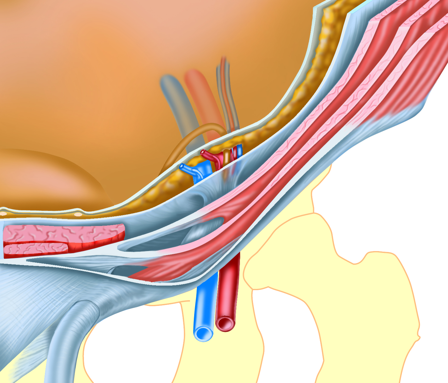
This image shows the abdominal wall layers forming the inguinal canal in a semi-3D exploded view. It can be seen how the inguinal canal traverses through and is formed by the layers of the abdominal wall. The internal and external inguinal ring can be seen, as well as the structures passing through the inguinal canal and froming the funiculus in the male. The space inferior of the overarching internal oblique and transverse abdominis muscle aponeuroses, through which a medial inguinal hernia can pass, is drawn exaggeratedly large to make clear the route of this hernia. This image originates from the e-learning lesson 'CASK inguinal area for students' at www.caskanatomy.info/inguinalarea created by the dept. of Anatomy and Embrylogy of Leiden University Medical Center, the Netherlands.
Anatomical structures in item:
Uploaded by: opgobee
Netherlands, Leiden – Leiden University Medical Center, Leiden University
Musculus obliquus externus abdominis
Musculus obliquus internus abdominis
Transversus
Fascia transversalis
Canalis inguinalis
Anulus inguinalis superficialis
Anulus inguinalis profundus
Peritoneum
Ductus deferens
Arteria testicularis
Vena testicularis
Funiculus
Fascia endoabdominalis
Arteria epigastrica inferior
Vena epigastrica inferior
Tendo conjunctivus
Inguen
Creator(s)/credit: S. Bas Blankevoort, medical illustrator, LUMC; O. Paul Gobée MD, anatomist and e-learning developer, LUMC
Requirements for usage
You are free to use this item if you follow the requirements of the license:  View license
View license If you use this item you should credit it as follows:
- For usage in print - copy and paste the line below:
- For digital usage (e.g. in PowerPoint, Impress, Word, Writer) - copy and paste the line below (optionally add the license icon):
"Abdominal wall layers of the inguinal canal, semi 3D exploded view, no labels" at AnatomyTOOL.org by S. Bas Blankevoort, LUMC and O. Paul Gobée, LUMC, license: Creative Commons Attribution-NonCommercial-ShareAlike
"Abdominal wall layers of the inguinal canal, semi 3D exploded view, no labels" by S. Bas Blankevoort, LUMC and O. Paul Gobée, LUMC, license: CC BY-NC-SA


Comments