nid: 63710
Additional formats:
None available
Description:
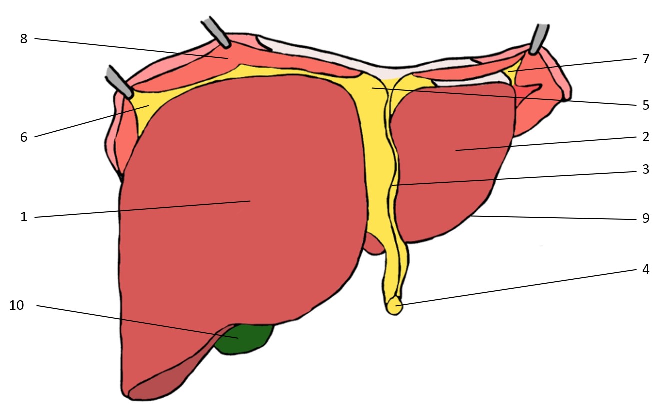
In this image we can study the diafragmatic surface (facies diafragmatica) of the liver as if it was taken out of the body by dividing it from its ligamentous attachments, the porta hepatis and the inferior vena cava.
Labels: Lobus dexter hepatis (1), Lobus sinister hepatis (2), Ligamentum falciforme hepatis (3), Ligamentum teres hepatis (4), Ligamentum coronarium hepatis (5), Ligamentum triangulare dextrum hepatis (6), Ligamentum triangulare sinistrum hepatis (7), Diafragma (8), Margo inferior hepatis (9), Vesica biliaris (10)
Universiteit Gent - Students - Drawing Hepar/Liver (2) - numbered Latin labels © 2023 by Manon Sierens is licensed under CC BY-NC-SA 4.0
Anatomical structures in item:
Uploaded by: sehellin
Belgium, Gent (Ghent) - Ghent University
Lobus hepatis dexter
Lobus hepatis sinister
Ligamentum falciforme hepatis
Ligamentum teres hepatis
Ligamentum coronarium hepatis
Ligamentum triangulare dextrum hepatis
Ligamentum triangulare sinistrum hepatis
Diaphragma
Margo inferior hepatis
Vesica biliaris (Fellea)
Creator(s)/credit: Manon Sierens, student, Belgium, Gent (Ghent) - Ghent University; Jana Foulon, student, Belgium, Gent (Ghent) - Ghent University; Senne Hellinck, student, Belgium, Gent (Ghent) - Ghent University
Requirements for usage
You are free to use this item if you follow the requirements of the license:  View license
 View license
 If you use this item you should credit it as follows:
- For usage in print - copy and paste the line below:
- For digital usage (e.g. in PowerPoint, Impress, Word, Writer) - copy and paste the line below (optionally add the license icon):
"Universiteit Gent - Students - Drawing Hepar/Liver (2) - numbered Latin labels" at AnatomyTOOL.org by Manon Sierens, Belgium, Gent (Ghent) - Ghent University, Jana Foulon, Belgium, Gent (Ghent) - Ghent University and Senne Hellinck, Belgium, Gent (Ghent) - Ghent University, license: Creative Commons Attribution-NonCommercial-ShareAlike
"Universiteit Gent - Students - Drawing Hepar/Liver (2) - numbered Latin labels" by Manon Sierens, Belgium, Gent (Ghent) - Ghent University, Jana Foulon, Belgium, Gent (Ghent) - Ghent University and Senne Hellinck, Belgium, Gent (Ghent) - Ghent University, license: CC BY-NC-SA


Comments