nid: 63534
Additional formats:
- Plate 8 with labels from Pelvis plastination specimens by Kees Maas, LUMC dept. Anatomy A4 CC version.pptx, *.pptx, 5MB, Powerpoint version with labels, for editing
Description:
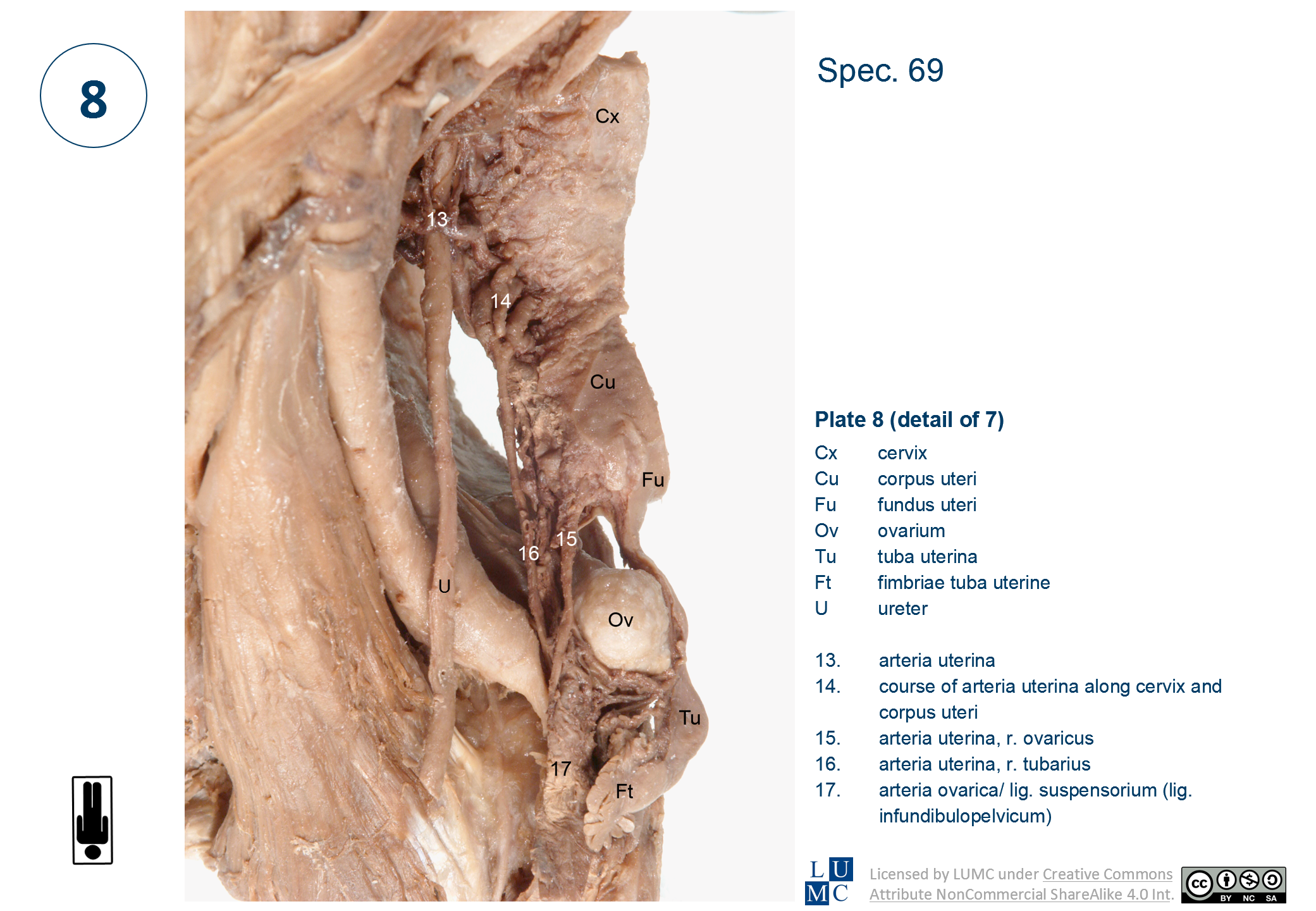
Frontal view of the female pelvis. The specimen shows details of the vascularisation of the uterus and ovary, with the anastomoses along the uterus between the vascularisation areas of uterus and ovary. Number labels with legend.
Anatomical structures in item:
Uploaded by: admin
Netherlands, Leiden – Leiden University Medical Center, Leiden University
Pelvis
Cervix uteri
Corpus uteri
Fundus uteri
Ovarium
Tuba uterina (Salpinx)
Fimbriae tubae uterinae
Ureter
Arteria uterina
Ramus tubarius (Arteria uterina)
Rami ovaricus (Arteria uterina)
Arteria ovarica
Ligamentum suspensorium ovarii
Creator(s)/credit: Kees (C.P.) Maas MD, PhD, dissection, LUMC; Prof. Marco C. DeRuiter PhD, anatomist, professor of Clinical and Experimental Anatomy, LUMC; J. Lens, medical photographer
Requirements for usage
You are free to use this item if you follow the requirements of the license:  View license
 View license
 If you use this item you should credit it as follows:
- For usage in print - copy and paste the line below:
- For digital usage (e.g. in PowerPoint, Impress, Word, Writer) - copy and paste the line below (optionally add the license icon):
"Leiden, Maas Photo 8 - Detail view of arteries of uterus and ovary (plastination specimen) - labels with legend" at AnatomyTOOL.org by Kees (C.P.) Maas, LUMC, Marco C. DeRuiter, LUMC and J. Lens, license: Creative Commons Attribution-NonCommercial-ShareAlike
"Leiden, Maas Photo 8 - Detail view of arteries of uterus and ovary (plastination specimen) - labels with legend" by Kees (C.P.) Maas, LUMC, Marco C. DeRuiter, LUMC and J. Lens, license: CC BY-NC-SA


Comments